The human body is marvelous in a lot of ways, but at the same time it can be a source of fear, discomfort and existential dread. Fascination and horror can really go hand in hand at times, but some folks make it their mission to learn and document what they can about our physical forms.
The “Medicalpedia” Instagram page is dedicated to sharing some of the more interesting and extreme cases of what can happen to a human body. Be warned, some of the images here and on the IG page are graphic. So settle in as you scroll through, upvote your favorites and be sure to share your thoughts in the comments below.
#1 Conjoined Twins Jadon And Anias, Fused At The Cranium, Were Successfully Separated After A 27 Hour Surgery Back In 2017
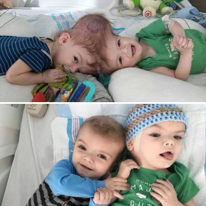
Image credits: medicalpedia
#2 Epidermal Burn Of The Hand Exposes Bright Colors Of Tattoo Ink Embedded In The Dermal Layer Of The Skin
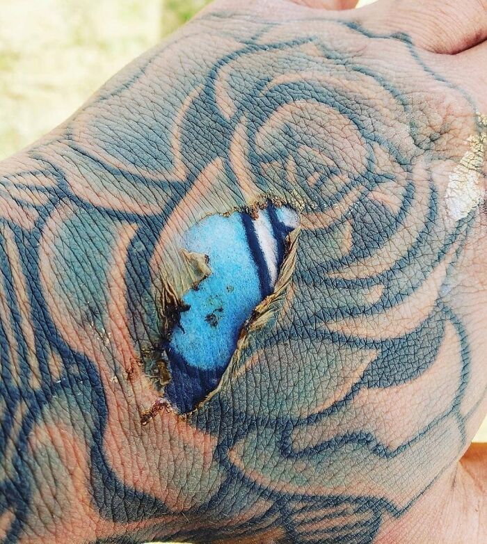
Image credits: medicalpedia
#3 Extensive Head Swelling Due To An Allergic Reation To Hair Dye
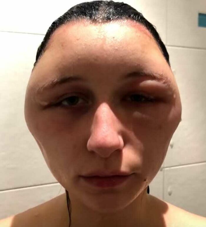
Image credits: medicalpedia
#4 A Graphic Comparison Between Healthy Lungs And Those Of A Heavy Smoker
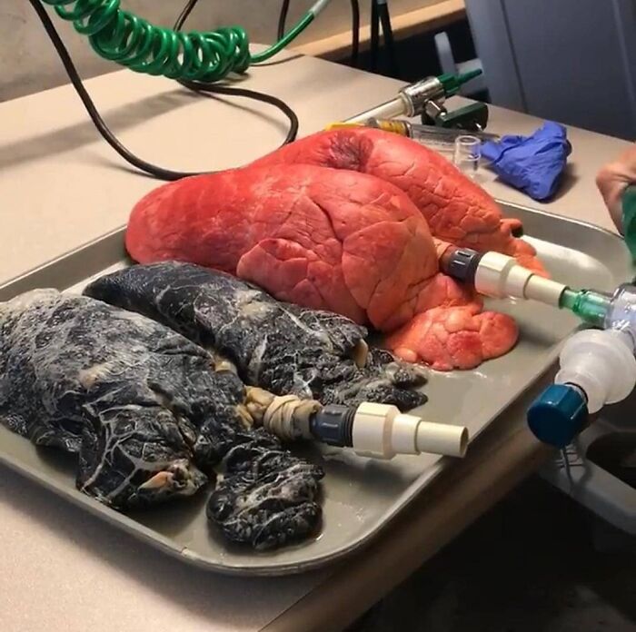
Image credits: medicalpedia
#5 Rare Case Of A Child Suffering From A Condition Called Hyperdontia
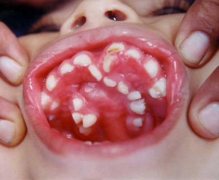
Image credits: medicalpedia
#6 Case Of Craniofacial Fibrous Dysplasia
Craniofacial fibrous dysplasia is a bone disease of the face and skull that replaces normal bone with fibrous-type tissue. This tissue is not as hard as normal bone, and because it is soft and stringy, it makes the bone more fragile and prone to break. Craniofacial fibrous dysplasia may cause shifting of facial features and facial asymmetry, such as incorrect placement of the eyes, misalignment of the jaw, and other problems. Fibrous dysplasia may appear in childhood, usually between the ages of 3 and 15. Boys are more often affected. Surgical treatment is usually required
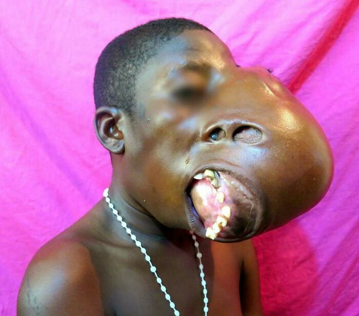
Image credits: medicalpedia
#7 Priceless Reaction Of A Dad Being Present At Birth

Image credits: medicalpedia
#8 Turning One Foot Backward To Go Forward:
Child cancer patient underwent an unusual surgery to reconstruct her leg after being diagnosed with bone cancer. The procedure is called rotationplasty and is done by removing the part of the femur that has cancer in it and dissecting the nerves and vessels away from the tissue. Then they rotate the bottom portion of the leg 180 degrees and attach it to the hip, bundling up the nerves and vessels so they are secure. The procedure is most commonly used to transfer the ankle joint to the knee joint following resection of a distal femoral bone tumor, such as osteosarcoma. The limb is rotated because the ankle flexes in the opposite direction compared to the knee. The benefit to the patient is that they have a functioning knee joint which gives a stable foundation for the patient to use a prosthetic leg successfully. Compared with endoprostheses, allografts, and amputations, rotationplasty patients have been reported to achieve superior functional outcomes in several studies. Despite this, rotationplasty is rarely performed owing to concerns with the psychological effect of the abnormal cosmetic appearance of the limb.
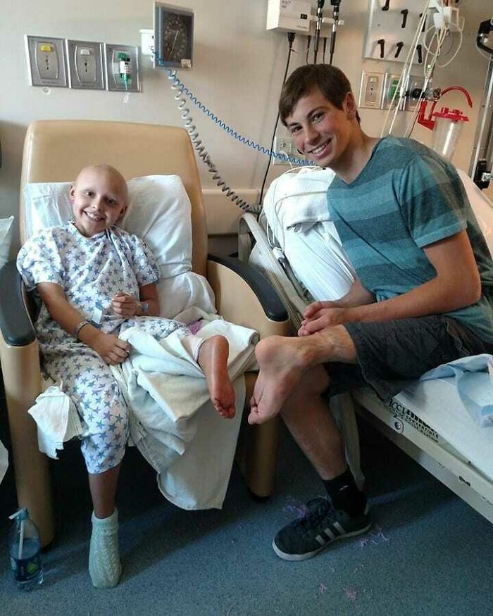
Image credits: medicalpedia
#9 Here Is An Interesting Case Of An Intraoral Presence Of Hair
A 61-year-old male came to the department complaining of discomfort in his mouth which has been present for six months. History revealed that the patient had a history of diagnosed and treated cancer of the buccal mucosa. He had been treated with radiation therapy and local resection was done along with surgical reconstruction with radial forearm flaps. Intraoral examination showed various hair fibers growing from the surface of the left buccal mucosa into the oral cavity and extending into the palate. Patient was advised to have laser excision of these hairs but he refused the procedure. His refusal was related to his previous surgical experiences. . Hairy intraoral flaps are one of rarest adverse effects of surgical reconstruction after treatment of oral malignancies. This may be due to the presence of hair follicles in the donor sites used as a flap. An intraoral hairy flap may result in constant discomfort affecting the quality of life.
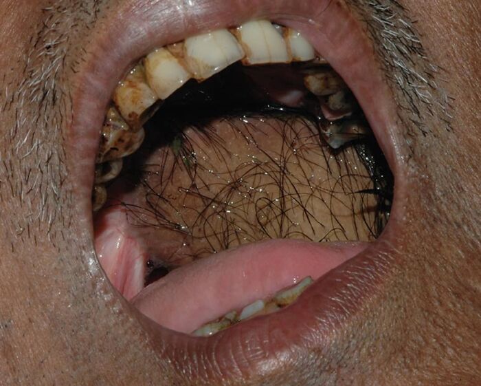
Image credits: medicalpedia
#10 Live Ladybug Found In A Man’s Transverse Colon During A Routine Colonoscopy
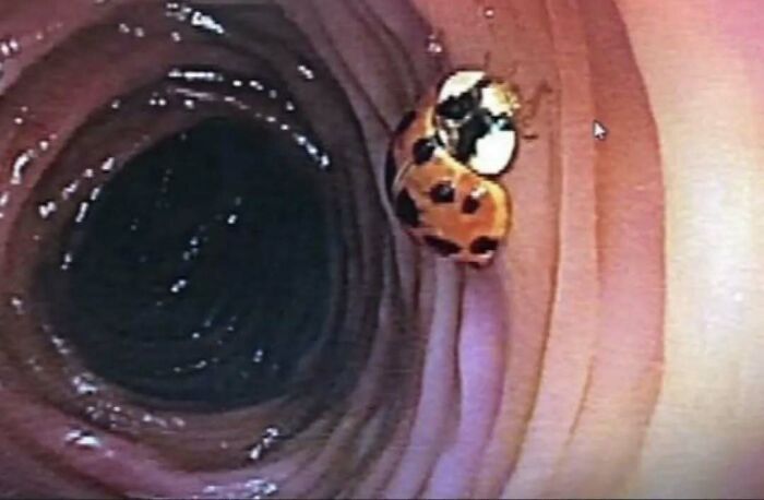
Image credits: medicalpedia
#11 When Dallas Wiens Was In His Early 20s, He Was Painting The Outside Of A Church When His Head Hit A High-Voltage Wire
Most of his face was burned off on contact; what was left was mostly destroyed through multiple surgeries to save his life. For two years, he lived without a face, with just a small opening where his mouth had once been. Then he qualified for a full facial transplant at Brigham & Women's Hospital — the first such surgery in the country. Now, after his 15-hour transplant, follow-up surgeries, and years of rehabilitation, Wiens has a full, rich life again. Severe electrical burns like Wiens suffered can lead to a loss of facial function and even facial features. Chemical burns, animal attacks, cancer, dog bites, and other terrifying accidents can leave people similarly disfigured. Until recently, there was little recourse beyond a series of partial reconstructions that would leave severe scars and deformities. Yet facial transplantation remains rare, in part because it is still very controversial. Despite the procedure's successes, there have been serious complications. There have also been some deaths due to post-surgery complications
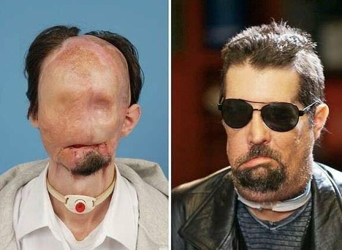
Image credits: medicalpedia
#12 Plicae Fimbriate - Ever Heard Of This Condition?
Plica fimbriata refers to the small folds in the membrane on the underside of your tongue. The folds tend to run parallel to, and on either side of your frenulum
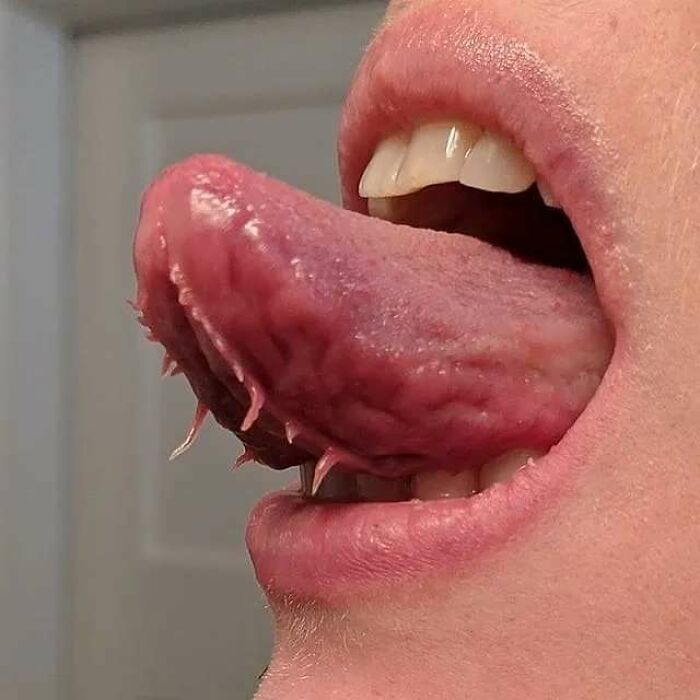
Image credits: medicalpedia
#13 26-Year Old Face Transplant Patient Reveals New Face
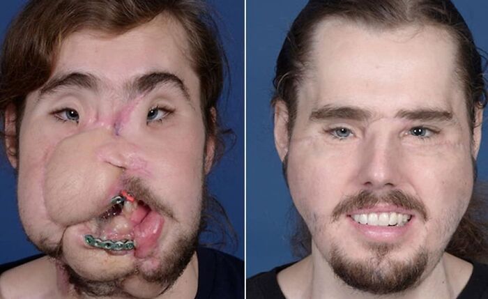
Image credits: medicalpedia
#14 Horrifying X-Ray Shows Why You Should Not Put Your Feet On Car Dashboards
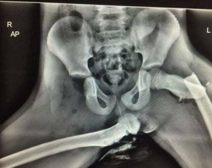
Image credits: medicalpedia
#15 Case Of A 17 Year Old Boy With An 18 Cm Long Tail
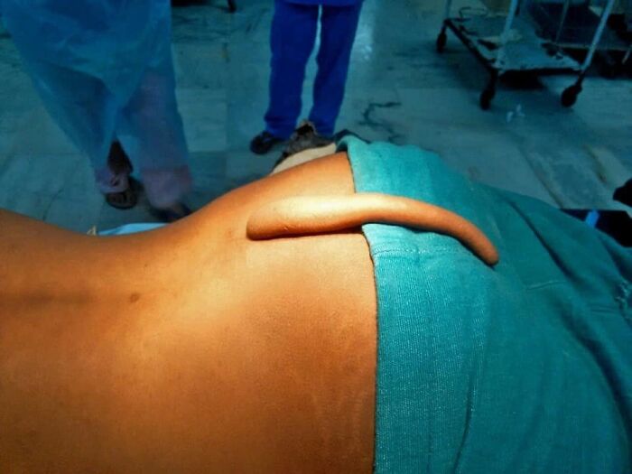
Image credits: medicalpedia
#16 Baby Encased In An Intact Amniotic Sac
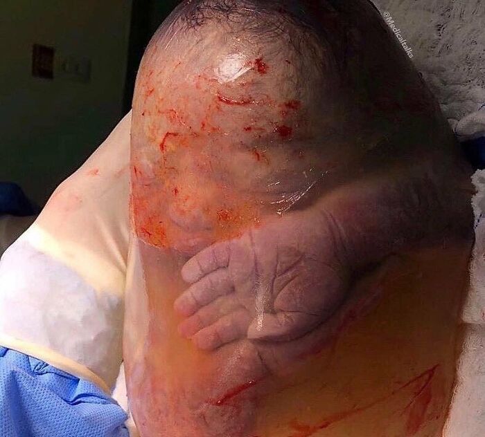
Image credits: medicalpedia
#17 Beautiful Close-Up Picture Of The Human Eye

Image credits: medicalpedia
#18 Car Crash Survivors Show ‘Scars’ From Seatbelts That Saved Their Lives
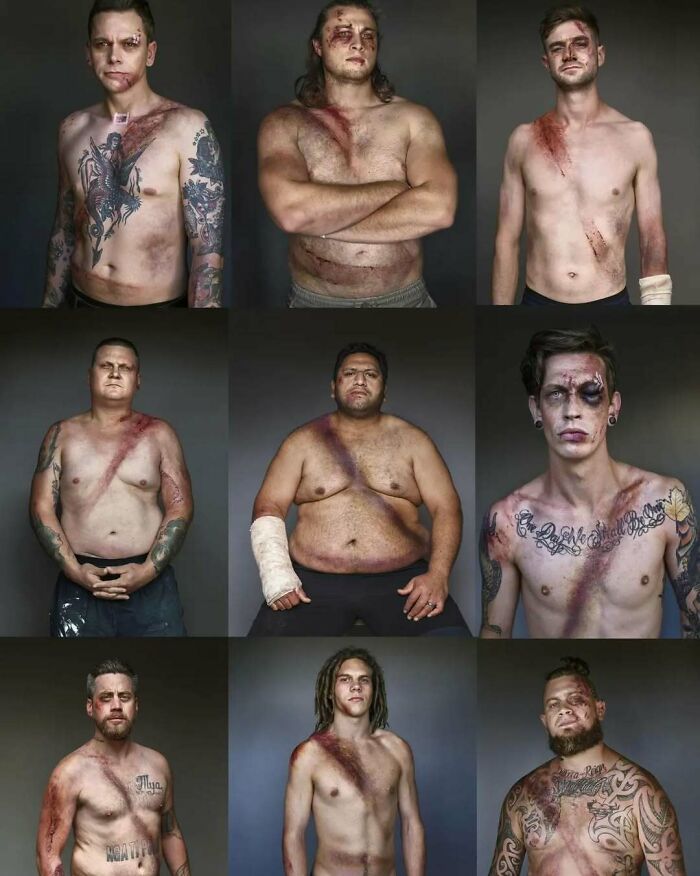
Image credits: medicalpedia
#19 Extracted Tooth With The Nerve Root Still Attached
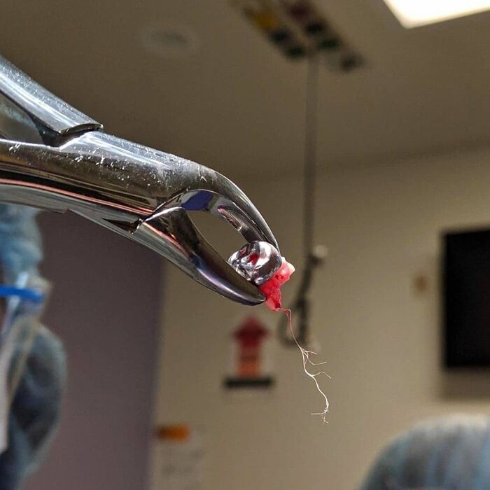
Image credits: medicalpedia
#20 Ever Wondered How A Foot Looks Like Under X-Ray While Wearing High Heels?
Studies have shown that these towering shoes can be costly in more ways than one, taking their toll on your spine, hips, knees, ankles and feet, while altering your posture and gait having harmful effects on the musculoskeletal system
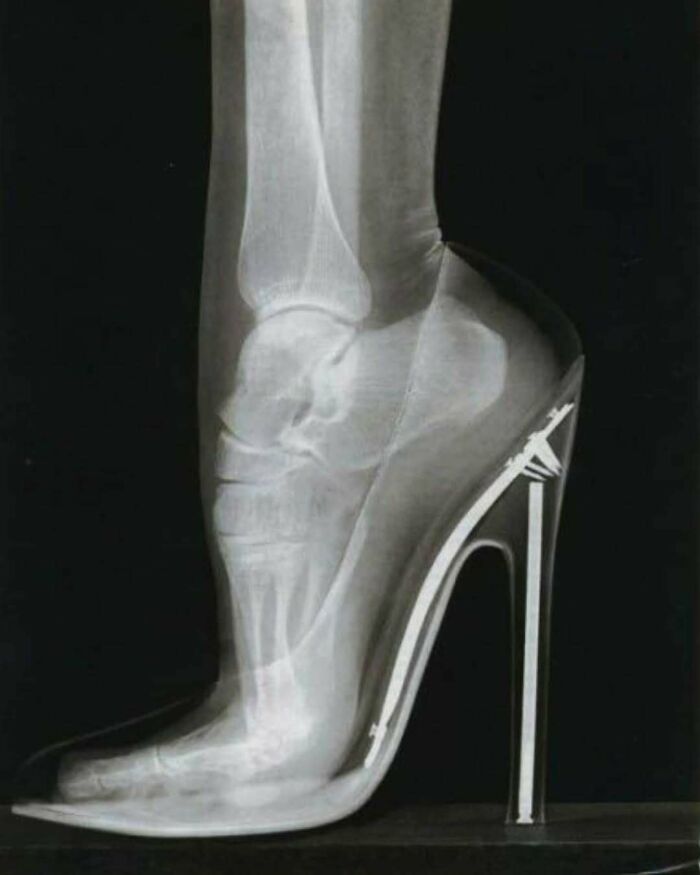
Image credits: medicalpedia
#21 Hair Growing Out Of Patient's Eye
Young male presented with a gradually increasing mass in the right eye since childhood. Examination revealed an elevated yellowish white lesion extending from 7 to 9 o’clock along the limbus with overlying fine vessels. Two hair follicles protruded from it. The clinical features were typical of limbal dermoid. The mass was excised with lamellar keratectomy, and histopathology further confirmed the diagnosis. Limbal dermoids are choristomas and are benign in nature. They may contain abberant tissues such as hair, teeth, bone, or muscle. Surgical excision is sometimes indicated for cosmetic purposes.

Image credits: medicalpedia
#22 The Human Body Has More Than 60,000 Miles Of Blood Vessels
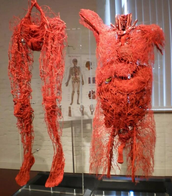
Image credits: medicalpedia
#23 Case Of A 13-Year Old Girl With Severe Scoliosis
This 13-year-old’s scoliosis was progressing so rapidly that major spinal surgery was her only treatment option. In just over six months, her curve progressed from what was initially 49-degree to a 99-degree curve. The girl now has a combination of titanium rods and screws around her spine. Luckily she fully recovered and got back to her normal activities. Scoliosis is a sideways curvature of the spine that occurs most often during the growth spurt just before puberty. While scoliosis can be caused by conditions such as cerebral palsy and muscular dystrophy, the cause of most scoliosis is unknown. About 3% of adolescents have scoliosis. Treatment depends on the degree of curve, location, and cause. Minor curves may simply be watched periodically.Treatments may include bracing, specific exercises, and surgery. The brace must be fitted to the person and used daily until growing stops. Specific exercises may be used to try to decrease the risk of worsening. They may be done alone or along with other treatments such as bracing. Evidence that chiropractic manipulation, dietary supplements, or exercises can prevent the condition from worsening is weak. However, exercise is still recommended due to its other health benefits. Surgery is usually recommended by orthopedists for curves with a high likelihood of progression (i.e., greater than 45 to 50° of magnitude), curves that would be cosmetically unacceptable as an adult, curves in people with spina bifida and cerebral palsy that interfere with sitting and care, and curves that affect physiological functions such as breathing. To completely straighten a scoliotic spine is usually impossible, but for the most part, significant corrections are achieved.
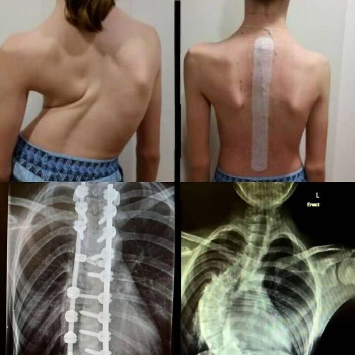
Image credits: medicalpedia
#24 An Extremely Rare Case Of Polydactyly Of The Toes With Synpolydactyly Of The Fingers
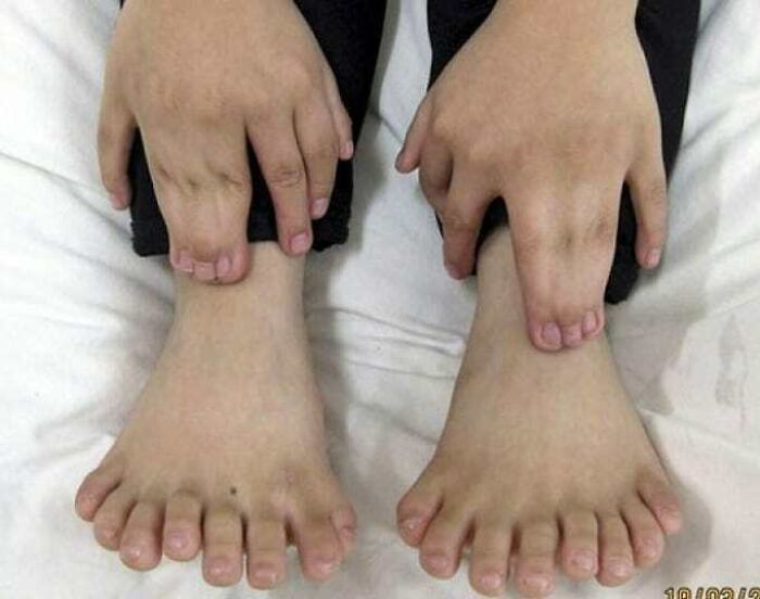
Image credits: medicalpedia
#25 Amputated Index Finger Turned Into A Keychain
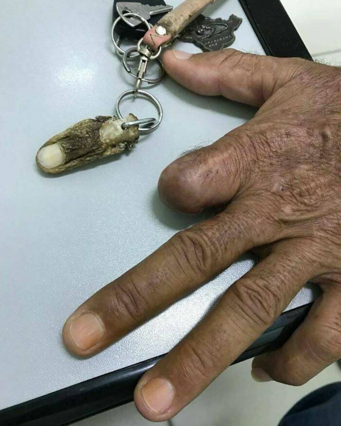
Image credits: medicalpedia
#26 Intact Blood Clot That Was Attached To The Ett (Endotracheal Tube) Of A Terminally Extubated Covid Patient
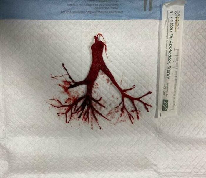
Image credits: medicalpedia
#27 Picture Of A 5-Year-Old Girl Comforting And Supporting Her 4-Year-Old Brother With Leukemia Struggling With The Side Effects Of Chemotherapy

Image credits: medicalpedia
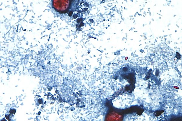
#28 Charcoaled Lungs Of A Heavy Smoker!
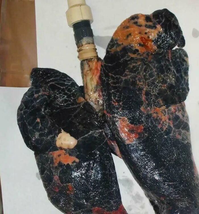
Image credits: medicalpedia
#29 A 31 Year Old Chinese Man, Stung By Multiple Tentacle Box Jellyfish At Chawang Beach On Samui Island, Thailand
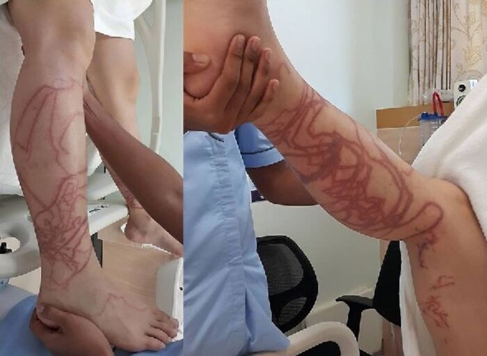
Image credits: medicalpedia
#30 This Is A Case Of A Lawn Mower Accident That Ended Up In Amputation Of The First And Second Toe
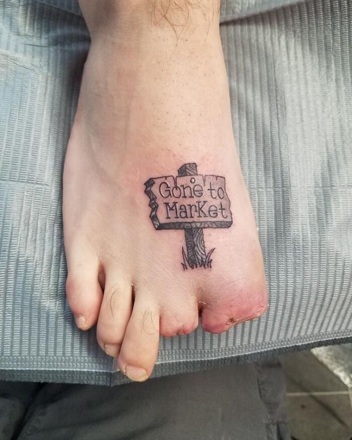
Image credits: medicalpedia
#31 This Is How They Used To Treat Headaches Back In 1850
It Was Believed That The Waves Generated From The Strike Relieved The Pain. A Century Later, It Was Proven That Those Waves Actually Sharpen The Pain And Make It More Intense.

Image credits: medicalpedia
#32 Ulnar Dimelia Or Mirror Hand Syndrome Is A Rare Congenital Anomaly Of The Upper Limb Characterized By Absence Of Radius, Duplication Of Ulna And Symmetric Polydactyly

Image credits: medicalpedia
#33 X-Ray Of A Child With A Disease Known As Osteogenesis Imperfecta Type III With Progressive Deformities In The Lower Extremities, Together With Severe Osteoporosis, Fragile Bones, And Coxa Vara
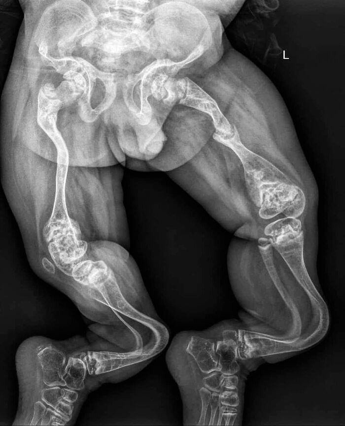
Image credits: medicalpedia
#34 A Peanut Lodged Inside A Child's Trachea!
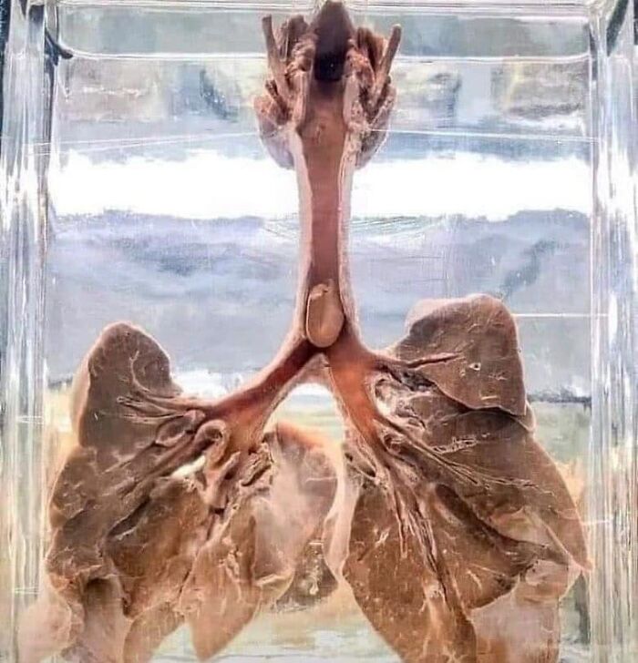
Image credits: medicalpedia
#35 Photo From 1950, Showing Iron Lungs In A Polio Ward
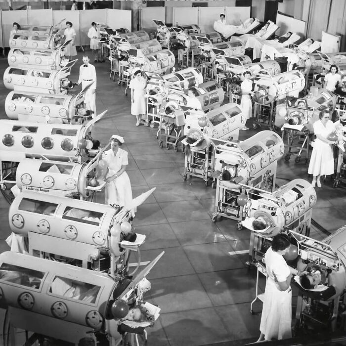
Image credits: medicalpedia
#36 The Biggest Baby By Natural Vaginal Birth Weighing Roughly 17.47 Pounds (7.9 Kg) And 22.5 Inches (57.5 Cm) Long
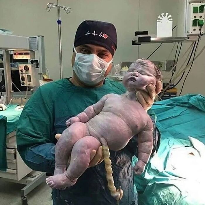
Image credits: medicalpedia
#37 Trauma Caused By An Agricultural Machine, Severing The Hand In Multiple Locations
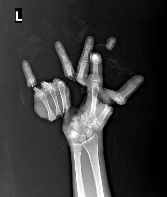
Image credits: medicalpedia
#38 Centers For Disease Control And Prevention (Cdc) Has Just Released A Case Study Of The First Pediatric Case Of Tetanus In Over 30 Years In The United States
A boy aged 6 years who had received no immunizations sustained a forehead laceration while playing outdoors on a farm; the wound was cleaned and sutured at home. Six days later, he had episodes of crying, jaw clenching, and involuntary upper extremity muscle spasms, followed by arching of the neck and back (opisthotonus) and generalized spasticity. Later that day, at the onset of breathing difficulty, the parents contacted emergency medical services, who air-transported him directly to a tertiary pediatric medical center. The boy subsequently received a diagnosis of tetanus and required approximately 8 weeks of inpatient care, followed by rehabilitation care, before he was able to resume normal activities. The boy required 57 days of inpatient acute care, including 47 days in the intensive care unit. The inpatient charges totaled $811,929 (excluding air transportation, inpatient rehabilitation, and ambulatory follow-up costs). One month after inpatient rehabilitation, he returned to all normal activities, including running and bicycling. Despite extensive review of the risks and benefits of tetanus vaccination by physicians, the family declined the second dose of DTaP and any other recommended immunizations. (photo is not the actual patient) Credit: Guzman-Cottrill JA, Lancioni C, Eriksson C, Cho Y, Liko J. Notes from the Field: Tetanus in an Unvaccinated Child
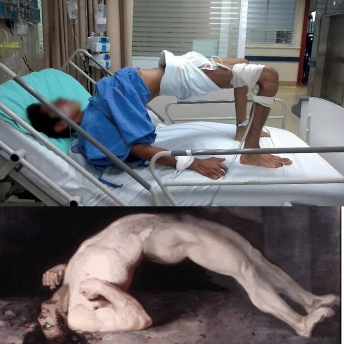
Image credits: medicalpedia
#39 Ant Heads Used Instead Of Sutures For Wound Closure. Because Of Their Strong Jaws, They Are Used As Emergency Sutures, When Nothing Else Is Available
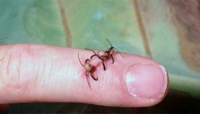
Image credits: medicalpedia
#40
The 27-Year-Old Woman With A History Of Schizoaffective Disorder Presented From An Inpatient Psychiatric Facility. Upon Entering The Stomach, There Were Numerous Crayons Layered In The Gastric Fundus And Gastric Body

Image credits: medicalpedia
#41 A 16-Year-Old Survied An Accident In Which A Spear Gun His Friend Was Holding Accidentally Discharged
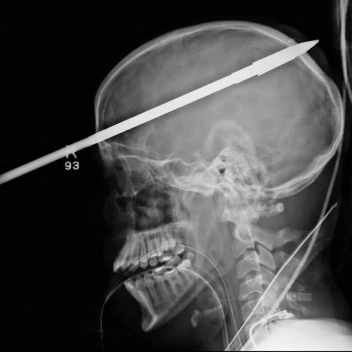
Image credits: medicalpedia
#42 Miracle Breast Implant Saves Woman's Life From Close-Range Gunshot Wound
A woman in Toronto, Canada, survived a gunshot wound to the chest thanks to her silicone breast implants, according to a case study published in the SAGE medical journal. The 30-year-old woman was shot at close range and walked into a local emergency department with a bullet wound on her left breast and a fractured rib on her right side. Surgeon who treated the patient, said the bullet entered the skin on the left side and then ricocheted across the patient's sternum into the right breast, breaking the rib. The breast implant likely deflected the bullet's trajectory, saving the patient's life, as the heart and lungs are located on the left side of the chest. The patient was treated by removing the implants, irrigating the wound, and prescribing antibiotics. The firearm was never recovered and the shooter remains unknown.
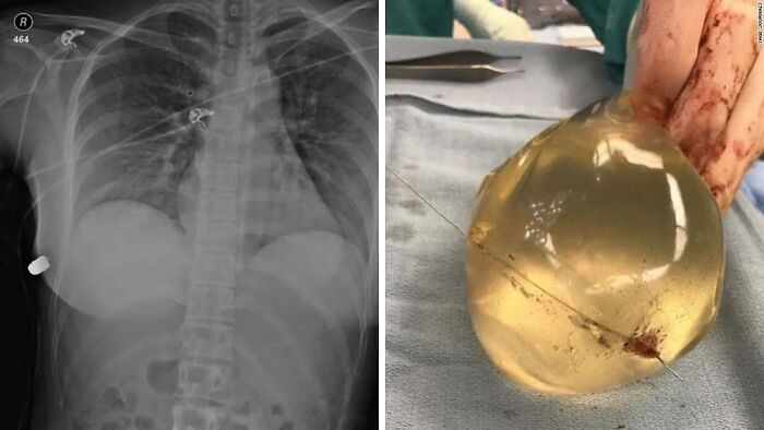
Image credits: medicalpedia
#43 An Interesting Case Of Triphalangeal Thumb
riphalangeal thumb (TPT) is a congenital hand anomaly in which the thumb has an additional phalanx. The true incidence of the condition is unknown, but is estimated at 1:25,000 live births. In about two-thirds of the patients with triphalangeal thumbs, there is a hereditary component. Clinical presentation of triphalangeal thumb can vary considerably and can be present in both hands or unilateral. The thumb can be long with a finger-like appearance. The presence of clinodactyly depends on the shape of the extra phalanx varying from wedge-shaped to rectangular. Various joints, ligaments, muscles, and tendons of the first ray can be hypoplastic or absent, with varying degrees of stiffness or instability. In general the surgical treatment is done for improvement of the thumb function. However, an extra advantage of the surgery is the improvement in appearance of the thumb. In the past, surgical treatment of the triphalangeal thumb was not indicated, but now it is generally agreed that operative treatment improves function and appearance. Because an operation was not indicated in the past, there’s still a population with an untreated triphalangeal thumb.
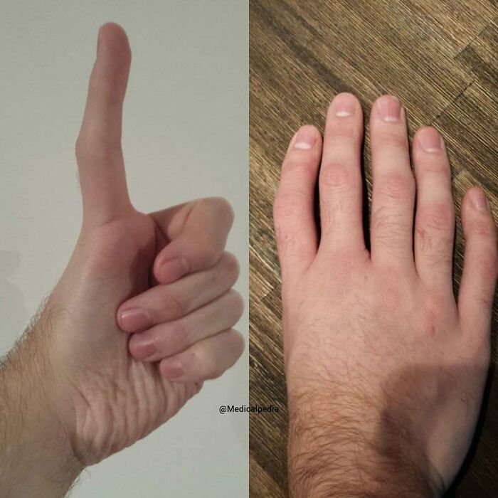
Image credits: medicalpedia
#44 Transillumination Of A Newborns Head, That Was Affected By Herpes Simplex Virus, Causing Severe Encephalitis
The infection was unfortunately not discovered in the hospital in time, resulting in destruction of 90% of his brain tissue. The light is showing the extent of damage. Herpes simplex encephalitis (HSE) is the most common single cause of viral encephalitis in infants and children. Treated or untreated, it can be associated with considerable morbidity and mortality, and its presentation is usually insidious and non-specific. Prompt and careful investigation is important in order to establish the diagnosis so that treatment can be optimised. Herpes simplex encephalitis is a devastating disease that can be difficult to diagnose in its early stages. By definition, neonatal herpes simplex (HSV) disease presents in the first 4 weeks of life and is almost always acquired by perinatal exposure to HSV. Illness symptoms often begin between the first 7–21 days of life. There is a spectrum of clinical syndromes; encephalitis is the most serious presentation, usually associated with lethargy, fever and convulsions. Identifying the source of HSE in neonatal disease can be difficult as a history of known maternal HSV disease (genital or occasionally oral) is not universal and maternal disease may be asymptomatic. Childhood HSE presents with similar features: fever, altered mental state (encephalopathy), a deteriorating level of consciousness, focal seizures or focal neurological abnormalities. The infective source is usually elusive. Parents can describe encephalopathy as a change in behaviour, sleepiness or confusion.

Image credits: medicalpedia
#45 The Hole That A Needle/Syringe Leaves In Your Skin Seen With An Electron Microscope
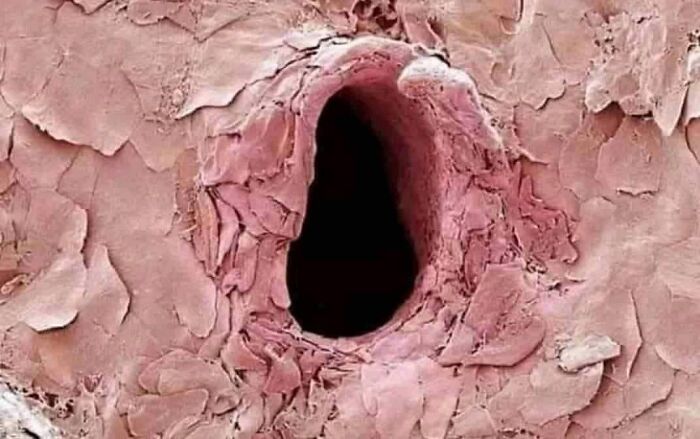
Image credits: medicalpedia
#46 When You're On A Stretcher But Just Gotta Have A Cigarette

Image credits: medicalpedia
#47 Dad Manages To Get A Crayon Embedded In His Foot!
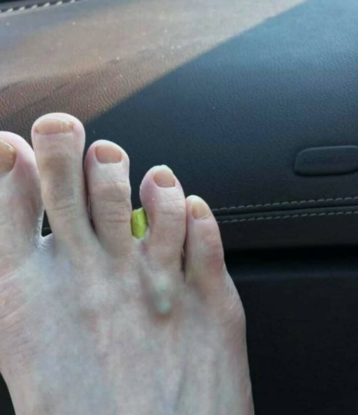
Image credits: medicalpedia
#48 A Case Of Raynaud's Syndrome Also Known As Raynaud's Phenomenon. It Is A Medical Condition In Which Spasm Of Arteries Cause Episodes Of Reduced Blood Flow
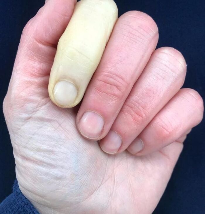
Image credits: medicalpedia
#49 Young Man Born With Fourteen Toes
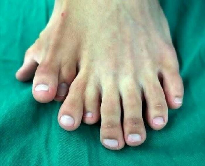
Image credits: medicalpedia
#50 In Case You Ever Wondered What Injecting 10ml Of Elemental Mercury Would Do To You
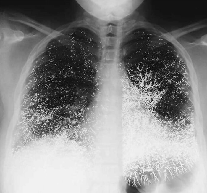
Image credits: medicalpedia
#51 Jugular Vein Distention, Which Is Quite Massive, In A Woman With Cardiac Tamponade
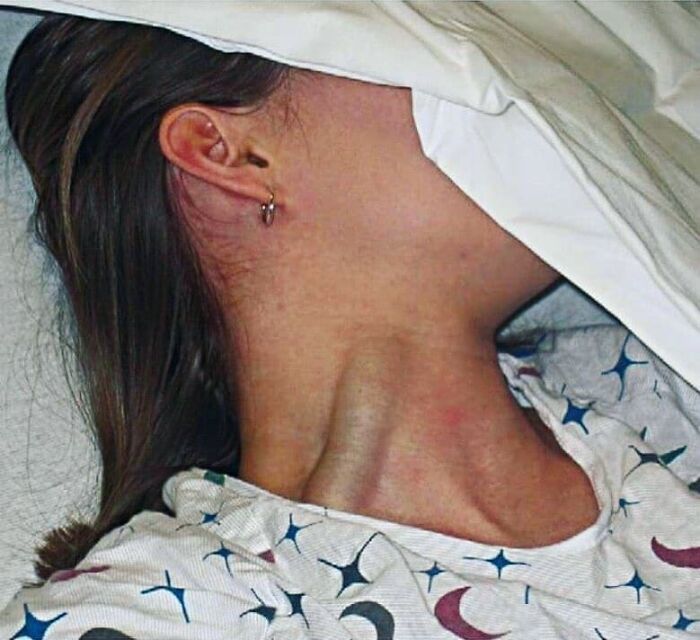
Image credits: medicalpedia
#52 Child’s Skull With A Part Of The Jaw Bone Being Removed To Show The Adult Teeth Developing Underneath The Deciduous Teeth (Baby Teeth)
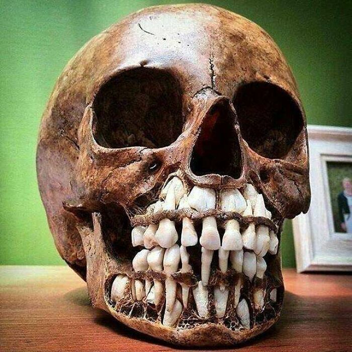
Image credits: medicalpedia
#53 Attempted S*****e With A Small Crossbow
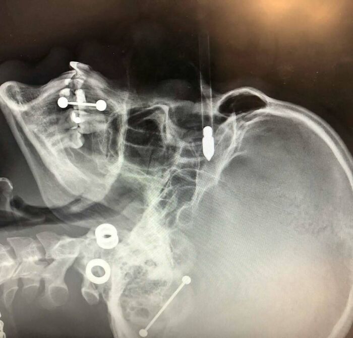
Image credits: medicalpedia
#54 A 15-Year-Old Boy Has Three Extra Vertebrae In His Neck
A Condition Known As Supernumerary Vertebra Along With Congenital Scoliosis - Causing Pain, Stress On His Nerves, And Making It Difficult For Him To Walk.
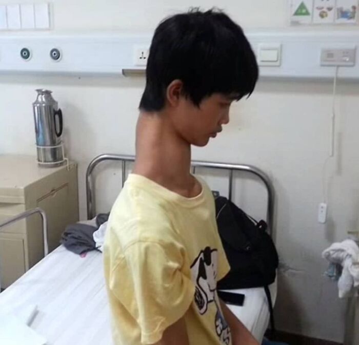
Image credits: medicalpedia
#55 X-Ray Of An Individual-A Contortionist In An Extreme Pose Of Spinal Extension
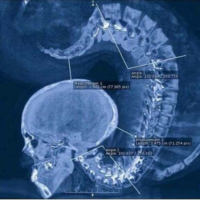
Image credits: medicalpedia
#56 This Woman Got Miraculously Pregnant With Quadruplets From An In-Vitro Fertilization! This Is Called A Monochorionic Diamniotic Quadruplet Pregnancy
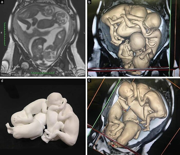
Image credits: medicalpedia
#57 Rugby League Accident Causinga Severe Arm Fracture
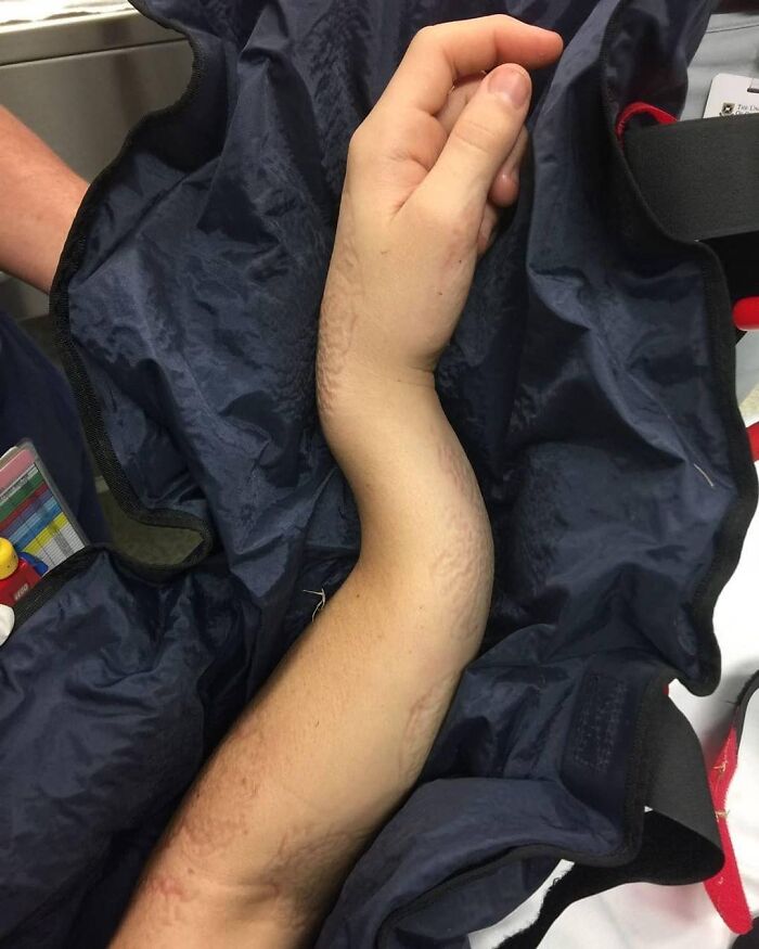
Image credits: medicalpedia
#58 A Condition Called Cvg
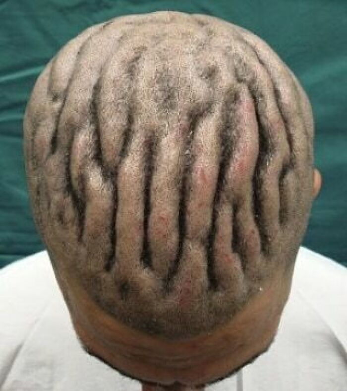
Image credits: medicalpedia
#59 Interesting Case Of A Child Having Red Eyes Due To Subconjuctival Hemorrhage
A subconjunctival hemorrhage occurs when a tiny blood vessel breaks just underneath the clear surface of your eye (conjunctiva). The conjunctiva can't absorb blood very quickly, so the blood gets trapped. You may not even realize you have a subconjunctival hemorrhage until you look in the mirror and notice the white part of your eye is bright red. A subconjunctival hemorrhage often occurs without any obvious harm to your eye. Even a strong sneeze or cough can cause a blood vessel to break in the eye. The most obvious sign of a subconjunctival hemorrhage is a bright red patch on the white (sclera) of your eye. Despite its bloody appearance, a subconjunctival hemorrhage should cause no change in your vision, no discharge from your eye and no pain. Your only discomfort may be a scratchy feeling on the surface of your eye

Image credits: medicalpedia
#60
Iris Freckles Are Tiny, Dark Brown Flecks On The Surface Of The Colored Part Of The Iris. This Changes In The Iris Suggest That The Person Could Have Been Overexposed To High Cumulative Doses Of Sunlight.
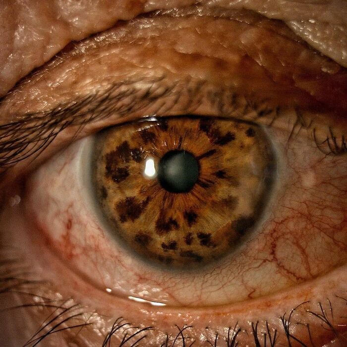
Image credits: medicalpedia
#61 Nineteen Year Old Bernard Underwent Reconstructive Surgery To Remove The Growth On His Face That Had Been There For More Than A Decade
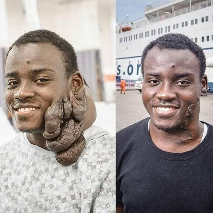
Image credits: medicalpedia
#62 It's A Photo Distorsion Caused By A Patient Moving In The Middle Of A Ct Scan Due To A Panic Attack As A Result Of Having Claustrophobia
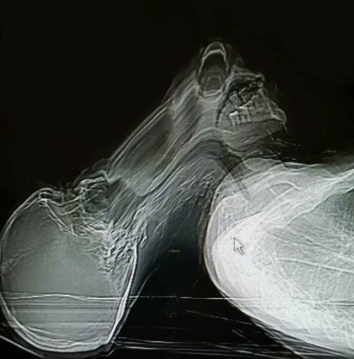
Image credits: medicalpedia
#63 X-Rays Of A 16-Month-Old Boy Who Swallowed A Spongebob Squarepants Necklace Pendant
Doctors who scanned a toddler's chest to try and discover what he had swallowed were shocked to find an image of the cartoon character SpongeBob SquarePants staring back at them. Doctor said the detail on the pendant was so clear because it was made up of tiny ridges in the metal's surface. The pendant was removed from the boy's throat and he was discharged to recover at home.
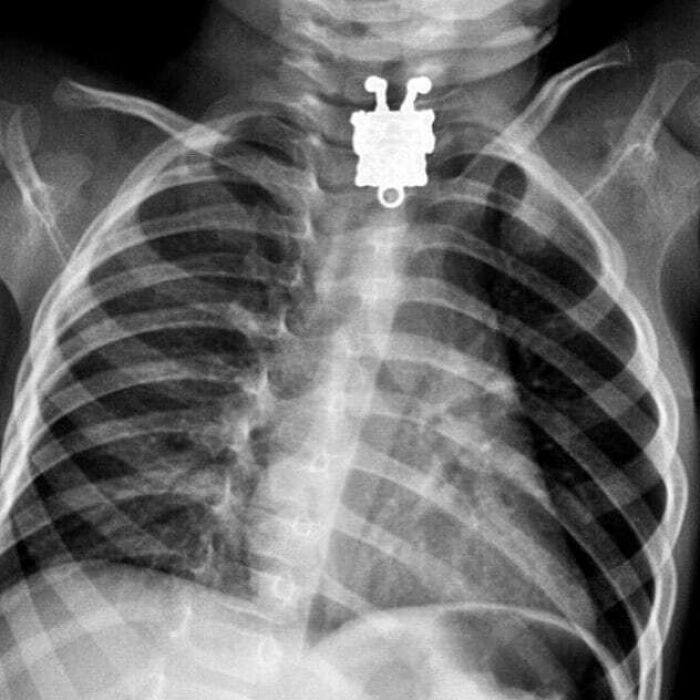
Image credits: medicalpedia
#64 Table Football Can Be A Dangerous Game!
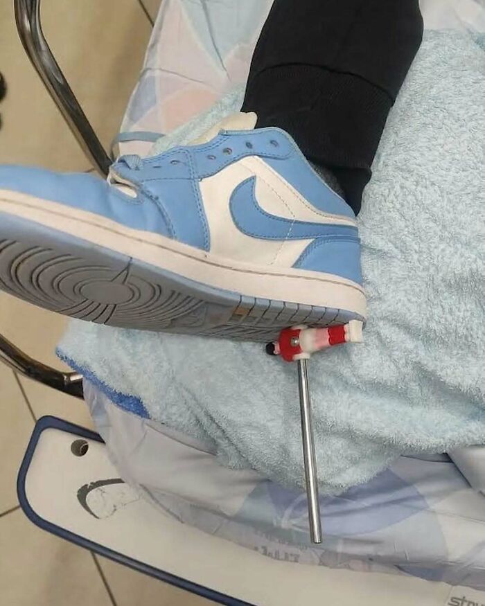
Image credits: medicalpedia
#65 Laparotomy Was Preformed To Remove The Foreign Body (Yankee Candle) Followed By A Temporary Colostomy
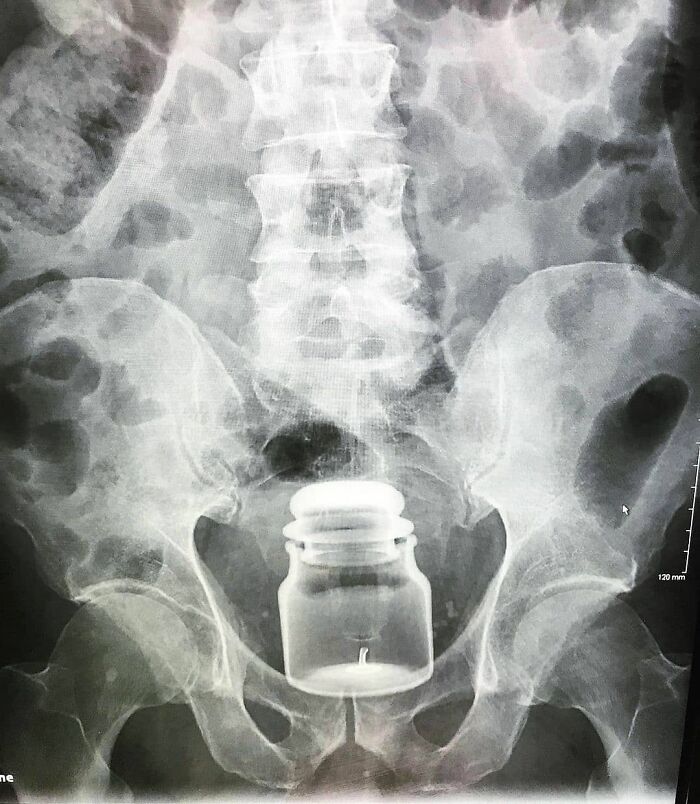
Image credits: medicalpedia
#66 This Is Arteriovenous Fistula Which Is A Surgically Made Connection Between An Artery And A Vein
You may ask why is this necessary and the answer is hemodialysis. An AV fistula causes extra pressure and extra blood to flow into the vein, making it grow large and strong. The larger vein provides easy, reliable access to blood vessels. Without this kind of access, regular hemodialysis sessions would not be possible. Untreated veins cannot withstand repeated needle insertions, because they would collapse the way a straw collapses under strong suction. At the start of a hemodialysis session, two needles are inserted into the vascular access. One needle carries blood from the body to the dialyzer, the other carries filtered blood back to the body. The blood then travels through a tube that takes it to the dialyzer. Inside the dialyzer, the blood flows through thin fibers that filter out wastes and extra fluid. The machine then returns the filtered blood to the body. A vascular access lets large amounts of blood flow continuously during hemodialysis treatments to filter as much blood as possible per treatment

Image credits: medicalpedia
#67 Syndactyly Is A Condition Wherein Two Or More Digits Are Fused Together
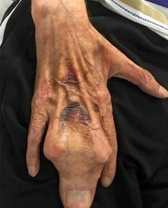
Image credits: medicalpedia
#68 Severe Case Of Teeth Calculus Build Up, Before And After Removal

Image credits: medicalpedia
#69 A Muay Thai Boxer Has Gone Viral After Images Emerged Of A Brutal Head Injury Sustained During A Fight
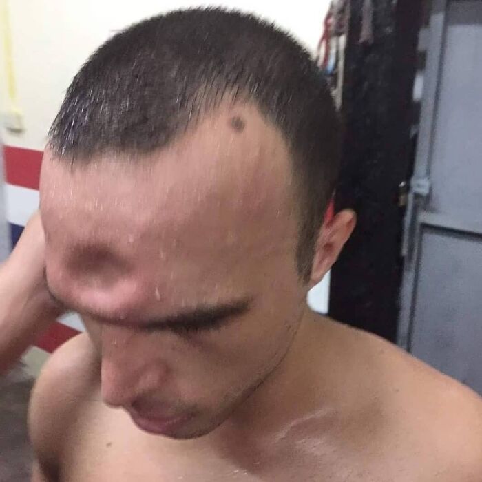
Image credits: medicalpedia
#70 Hey Nurse, I Think I Have Something Stuck In My Nose
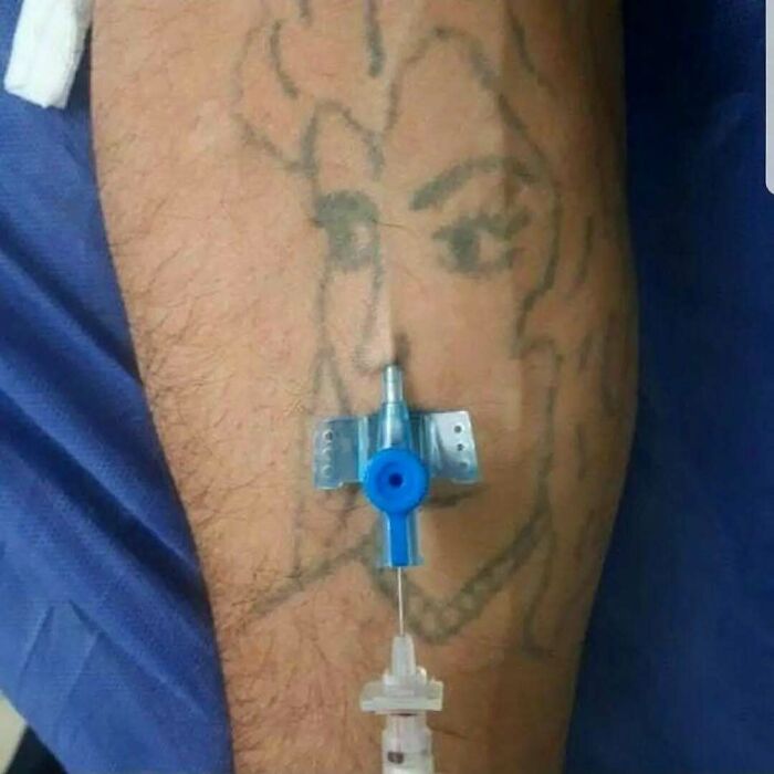
Image credits: medicalpedia
#71 Fascinating Comparison Between A Human And A Gorilla Skeleton

Image credits: medicalpedia
#72 Man Aged 36, Who Has Never Brushed His Teeth Since Childhood, Came With Severe Tooth Decay
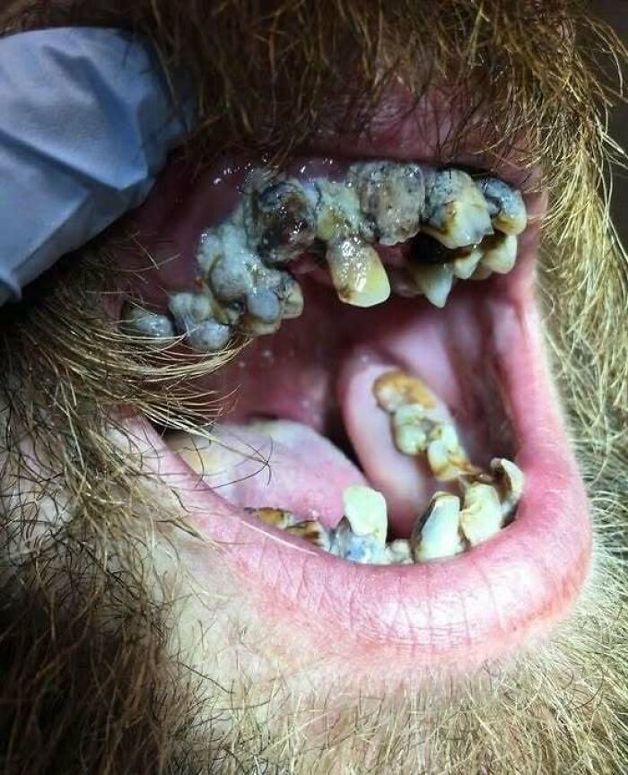
Image credits: medicalpedia
#73 A Ring Burried Underneath The Skin
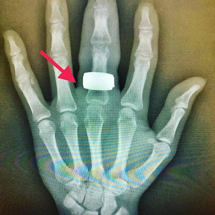
Image credits: medicalpedia
#74 Osteosarcoma Is The Most Common Histological Form Of Primary Bone Cancer
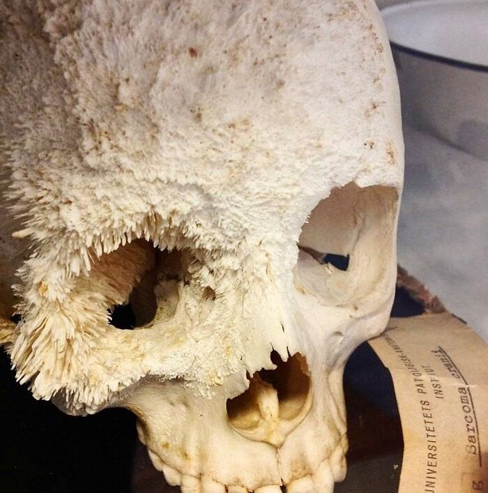
Image credits: medicalpedia
#75 A Patient Had Inserted In His Rectum Approximately 2 H Prior To Presentation
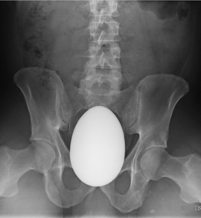
Image credits: medicalpedia
#76 Saved By A Plastic Bag! The Surprising Way To Help Premature Babies!
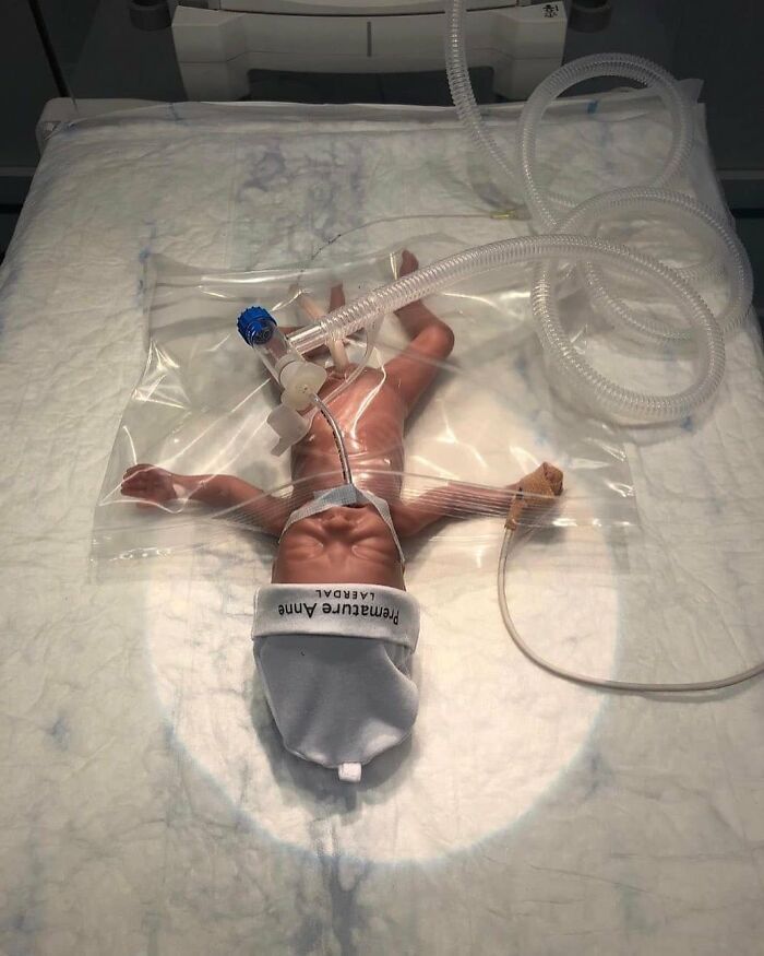
Image credits: medicalpedia
#77 Beautiful Photo Of Cerebellum
The cerebellum is one of the most identifiable parts of the brain due to its unique shape and location. It is located behind the top part of the brain stem and is made of two hemispheres (halves). It is extremely important for being able to perform everyday voluntary tasks such as walking and writing. It is also essential to being able to stay balanced and upright. Patients who have suffered from damaged cerebellums often struggle with keeping their balance and maintaining proper muscle coordination.
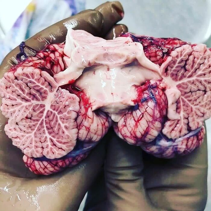
Image credits: medicalpedia
#78 An Interesting Photo, Showing How Dirt Doesn't Stick To The Scar
Can you guess why? A scar is an area of fibrous tissue that replaces normal skin after an injury. Scars result from the biological process of wound repair in the skin, as well as in other organs and tissues of the body. Because it’s a collagen matrix, it sometimes doesn’t have the same properties as normal skin thus not containing sweat glands and as a result, no dirt/dust sticks to it
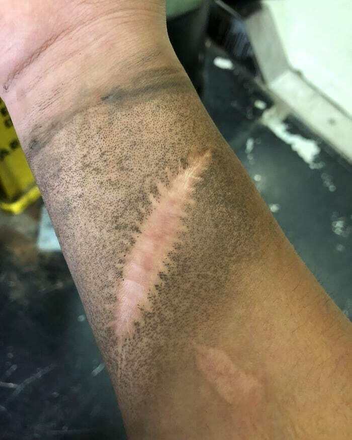
Image credits: medicalpedia
#79 Wwi Artillery Shell Stuck In Elderly Man's Rectum Causes Hospital Evacuation
An elderly man caused a stir at a hospital in Toulon, France when he arrived with a World War I artillery shell stuck in his rectum. The 88-year-old patient visited the hospital to have the antique explosive removed, but the hospital had to be partially evacuated and bomb disposal experts called in as a precaution. However, the experts determined that there was little chance of the shell exploding. The hospital then had to perform surgery to remove the object, which was almost 8 inches long and more than 2 inches wide. The patient is now recovering and is in good health.
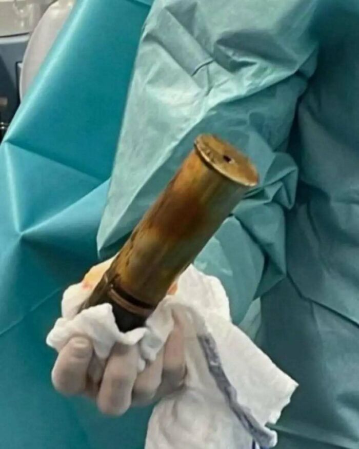
Image credits: medicalpedia
#80 Here's What You'd Look Like As Just A Nervous System!

Image credits: medicalpedia
#81 Removal Of A Fascinating Gallblader Stone
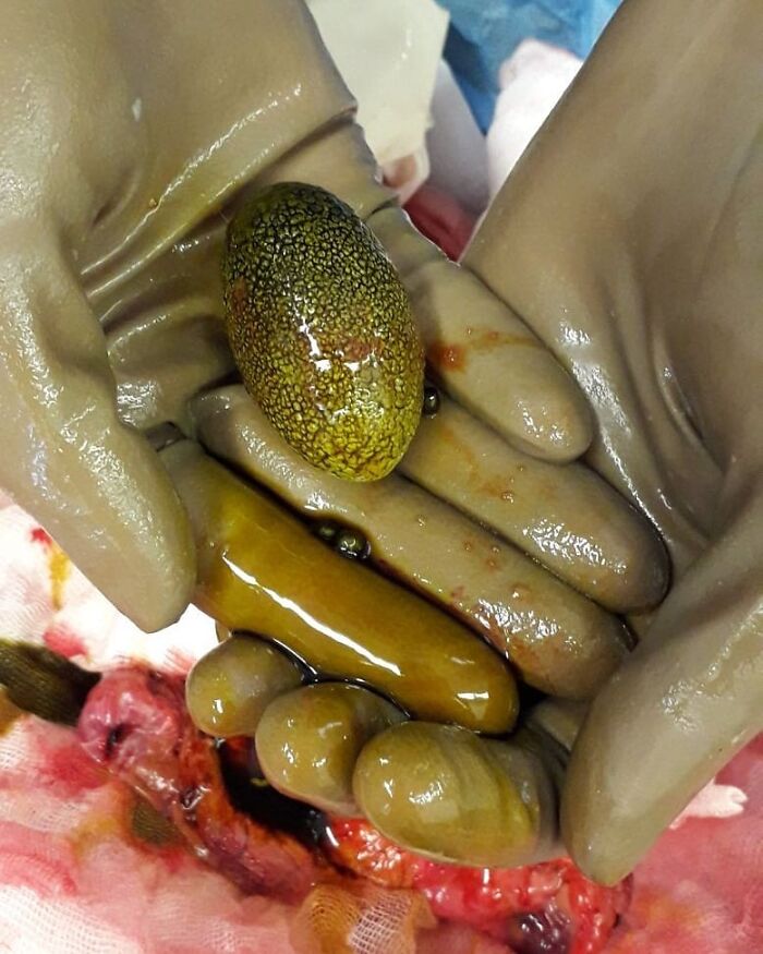
Image credits: medicalpedia
#82 This Is What Your Blood Looks Like During Holidays
This is a 4-ml sample of hyperlipidemic blood in a vacutainer with EDTA left to settle for four hours without centrifugation, the lipids separated into the top fraction. Hyperlipidemia is abnormally elevated levels of any or all lipids or lipoproteins in the blood. You can control some of its causes, but not all of them. Hyperlipidemia is treatable, but it's often a life-long condition. You'll need to watch what you eat and also exercise regularly. You might need to take a prescription medication, too. The goal is to lower the harmful cholesterol levels. Doing so reduces your risk of heart disease, heart attack, stroke, and other problems. Cholesterol, along with triglycerides and other fats, can build up inside your arteries. This makes the blood vessels narrower and makes it more difficult for blood to get through. Your blood pressure could go up. The buildup can also cause a blood clot to form. If a blood clot breaks off and travels to your heart, it causes a heart attack. If it goes to your brain, it can cause a stroke.

Image credits: medicalpedia
#83 Lightning Strike Causes Patterned Charring Along The Contact Points Of A Metallic Locket
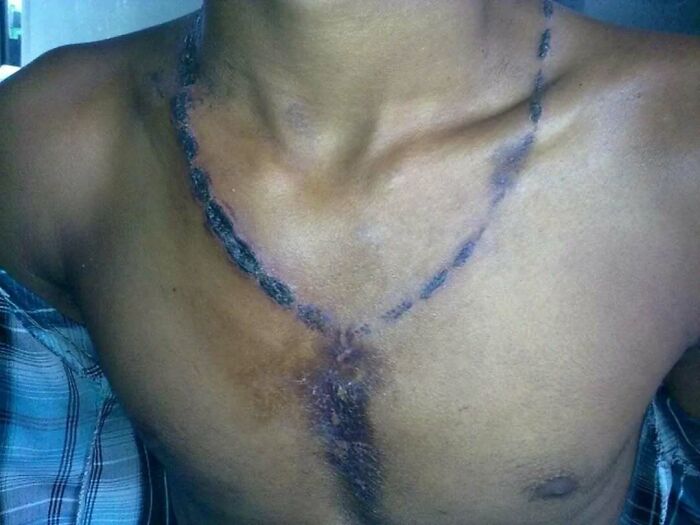
Image credits: medicalpedia
#84 The 24-Year-Old Has Injected His Arms For Four Years With A Substance Called Synthol As He Tried To Emulate The Cartoon Character's Muscles

Image credits: medicalpedia
#85 This 70-Year-Old Patient Never Brushed His Teeth In His Life!
The examination revealed existence of deposits on the teeth, known as calculus, and a lot of caries.Therapy included removal of deposits and multiple tooth extraction with dental prosthesis implantation. In dentistry, calculus or tartar is a form of hardened dental plaque. It is caused by precipitation of minerals from saliva and gingival crevicular fluid (GCF) in plaque on the teeth. This process of precipitation kills the bacterial cells within dental plaque, but the rough and hardened surface that is formed provides an ideal surface for further plaque formation. This leads to calculus buildup, which compromises the health of the gingiva (gums). Calculus formation is associated with a number of clinical manifestations, including bad breath, receding gums and chronically inflamed gingiva. Brushing and flossing can remove plaque from which calculus forms; however, once formed, calculus is too hard (firmly attached) to be removed with a toothbrush. Calculus buildup can be removed with ultrasonic tools or dental hand instruments
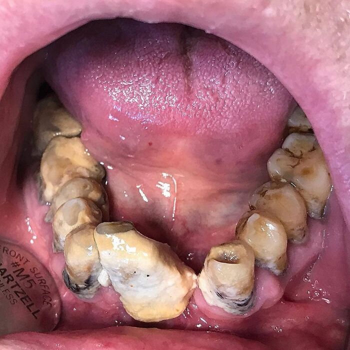
Image credits: medicalpedia
#86 For The Past Several Years A 31-Year-Old Man From India Had Been Watching The Tumor Emerge From The Back Of His Skull
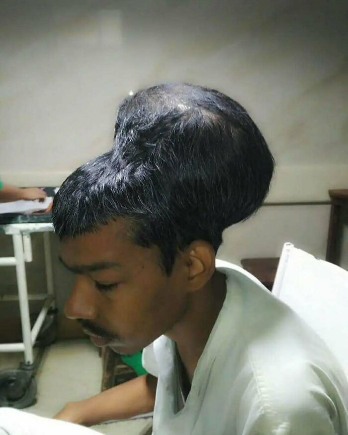
Image credits: medicalpedia
#87 An Unbelievable Case Of A Lightning Strike Survivor
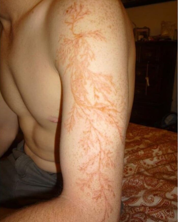
Image credits: medicalpedia
#88 Hand Washing Prior To Surgery
Do you know the history behind it? Ignaz Semmelweis, a Hungarian doctor working in Vienna General Hospital, is known as the father of hand hygiene. In 1846, he noticed that the women giving birth in the medical student/doctor-run maternity ward in his hospital were much more likely to develop a fever and die compared to the women giving birth in the adjacent midwife-run maternity ward. He decided to investigate, seeking differences between the two wards. He noticed that doctors and medical students often visited the maternity ward directly after performing an autopsy. Based on this observation, he developed a theory that those performing autopsies got ‘cadaverous particles’ on their hands, which they then carried from the autopsy room into the maternity ward. Midwives did not conduct surgery or autopsies, so they were not exposed to these particles. As a result, Semmelweis imposed a new rule mandating handwashing with chlorine for doctors. The rates of death in his maternity ward fell dramatically. This was the first proof that cleansing hands could prevent infection. However, the innovation was not popular with everyone: some doctors were disgruntled that Semmelweis was implying that they were to blame for the deaths and they stopped washing their hands, arguing in support of the prevailing notion at that time that water was the potential cause of disease. Sadly, the hand hygiene practices promoted by Semmelweis were not widely adopted. In general, handwashing promotion stood still for over a century. It was not until the 1980s, when a string of foodborne outbreaks and healthcare-associated infections led to public concern that the United States Centers for Disease Control and Prevention identified hand hygiene as an important way to prevent the spread of infection. In doing so, they heralded the first nationally endorsed hand hygiene guidelines, and many more have followed.
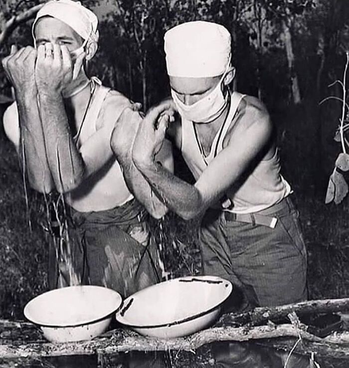
Image credits: medicalpedia
#89 Case Of A 87-Year-Old Woman With A Five Inch Cutaneous Horn
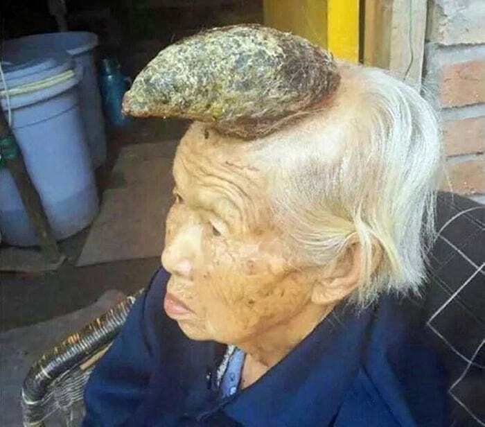
Image credits: medicalpedia
#90 A Case Of Heart-Shaped Posterior Synechiae Secondary To Uveitis
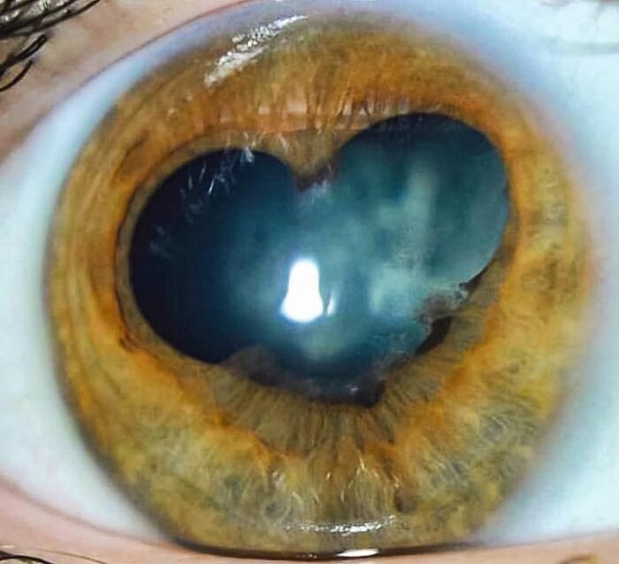
Image credits: medicalpedia
#91 An Interesting Case Of A Condition Called Arthritis Mutilans
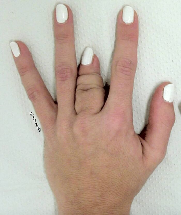
Image credits: medicalpedia
#92 X-Ray Photo Of A Patient Who Swallowed Spoons, Scewdrivers, Tootbrushes And A Knife!
A condition called acuphagia. This person SWALLOWED 2 screwdrivers, 2 toothbrushes, 8 spoons and a kitchen knife!! After a 4 hour surgery, all of the items were removed from his stomach(second picture) and he made a full recovery. Acuphagia is described as eating sharp metal objects and is recognised as a type of pica. Basically, pica refers to eating non-nutritive, non-food items in people without any underlying mental disorders. Pica is most commonly seen in pregnant women, small children and those with psychiatric disorders (developmental disorders, autism spectrum disorder, and even schizophrenia).
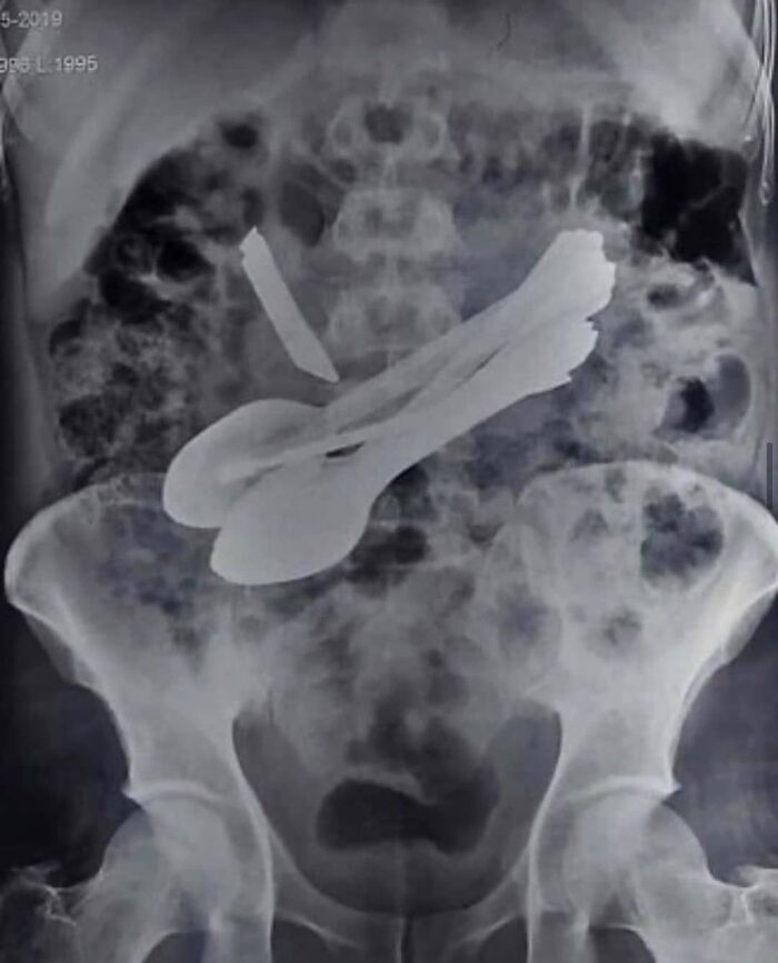
Image credits: medicalpedia
#93 Extreme Hypermobility Of The Finger
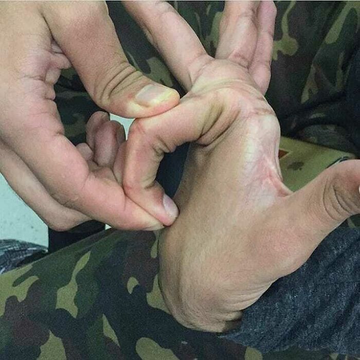
Image credits: medicalpedia

.png?w=600)