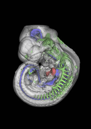
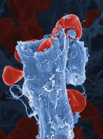
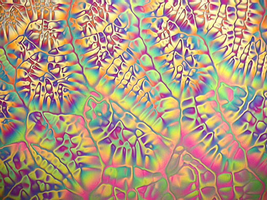
Special Award Winner: The new technique of Optical Projection Tomography enables stained whole embryos and small pieces of tissue to reveal internal structures without the need for cutting sections, as shown here on this mouse embryoPhotograph: James Sharpe/Wellcome TrustSpecial Winner: Crystals of oxidised Vitamin C (dehydroascorbic acid)Photograph: Spike Walker/Wellcome TrustSpecial Winner: Colour-enhanced image of red blood cells leaking from a ruptured blood vesselPhotograph: Anne Weston/Wellcome Trust
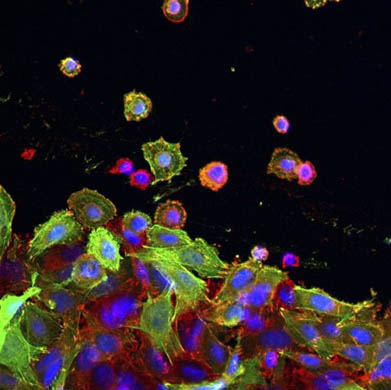
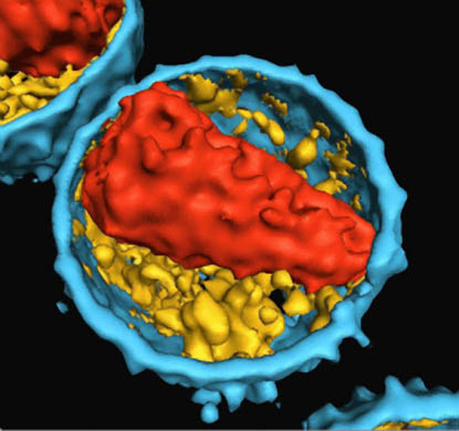
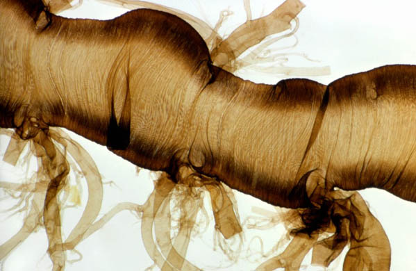
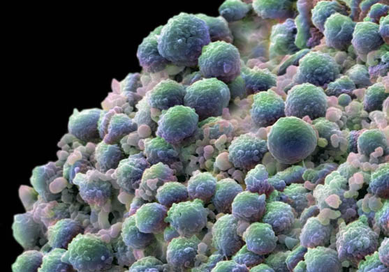
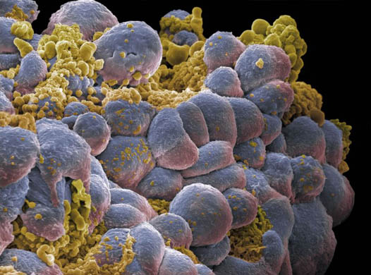
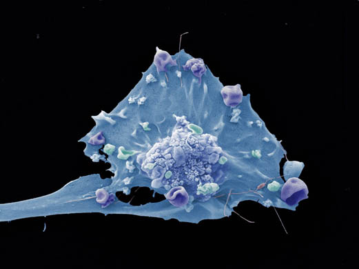
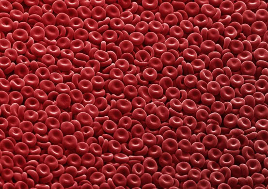
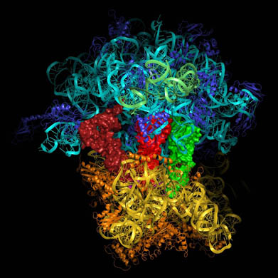
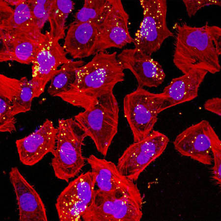
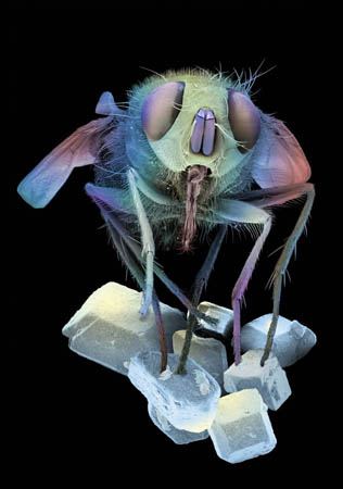
Villi from the human small intestinePhotograph: Stephanie Schuller/Wellcome TrustLiquid crystal seen under polarised lightPhotograph: Karen Neill/Wellcome TrustColon cancer cells growing in a dishPhotograph: Lorna McInroy/Wellcome TrustInternal structure of an HIV particlePhotograph: Stephen Fuller/Wellcome TrustTrachea from a silkwormPhotograph: Spike Walker/Wellcome TrustA clump of prostate cancer cells. The blue-green cells are growing, whereas the pink ones are dying by programmed cell death (apoptosis)Photograph: Dave McCarthy and Annie Cavanagh/Wellcome TrustA clump of breast cancer cells. The blue cells are actively growing whereas the yellow ones are in the process of dying by programmed cell death (apoptosis)Photograph: Dave McCarthy and Annie Cavanagh/Wellcome TrustColour-enhanced image of a single breast cancer cellPhotograph: Anne Weston/Wellcome TrustA carpet of red blood cells clearly showing their typical biconcave disc shapePhotograph: Dave McCarthy and Annie Cavanagh/Wellcome TrustA molecular model showing all the different molecules, both RNA (turquoise, green and yellow) and protein (purple and orange), that make up the ribosome, the RNA and protein found in all cellsPhotograph: Venki Ramakrishnan/Wellcome TrustMeningitis bacteriaPhotograph: Shao Jin Ong/Wellcome TrustA fly on sugar crystalsPhotograph: Dave McCarthy and Annie Cavanagh/Wellcome Trust
Sign up to read this article
Read news from 100’s of titles, curated specifically for you.