Scientists have believed for years that memories are strengthened during sleep through a conversation between the cortex, thalamus, and hippocampus. Direct evidence in humans has been almost impossible to obtain, because measuring it requires electrodes inside the brain.
Researchers at Kennedy Krieger Institute and Johns Hopkins Medicine have now done it. Using simultaneous intracranial recordings from the medial orbitofrontal cortex, thalamus, and hippocampus in 19 patients with epilepsy, they showed that slow oscillations in the orbitofrontal cortex organize the faster rhythms in all three regions during sleep, and that stronger coordination predicted better memory the next morning.
The work was published in the Proceedings of the National Academy of Sciences under the title "A hierarchical cascade of sleep rhythms supports motor memory and is hijacked by epileptic spikes in human epilepsy." The findings also showed that epileptic spikes disrupt the process.
Why This Could Only Be Studied This Way
The methodological constraint is the reason the study is described as a first, and it also defines its limits.
Scalp EEG records brain activity through the skull, which blurs signals and cannot reliably separate structures deep in the brain. The thalamus and hippocampus sit far from the surface. Measuring what they do simultaneously, at the timescale of individual oscillations, requires electrodes placed inside brain tissue.
That is not something researchers can do to healthy volunteers. It happens only when patients with drug-resistant epilepsy have electrodes implanted for clinical purposes, to identify precisely where their seizures begin before surgery. Electrode placement is determined entirely by clinical need.
The 19 subjects, covering 26 sampled hemispheres, were every patient admitted to the Massachusetts General Hospital epilepsy monitoring unit over a roughly two-and-a-half-year window whose stereo-EEG electrodes happened to sample all three target regions at once. That is a rare and narrow opportunity, and it is why the sample is 19 people rather than hundreds.
The Rhythms Form a Hierarchy
The recordings showed a nested structure rather than three regions simply firing together.
Three rhythms matter here. Slow oscillations are large, low-frequency waves during deep non-REM sleep. Sleep spindles are brief bursts of faster activity generated by the thalamus. Ripples are very fast, short events associated with the replay of recent experience in the hippocampus.
The researchers found that orbitofrontal slow oscillations robustly modulate both spindle and ripple activity, within their own region and across the thalamus and hippocampus. The slow waves act as a conductor, setting the timing that the faster rhythms follow.
Participants performed a validated motor learning task before sleeping, and their performance was retested afterward. Most combinations of oscillation rates positively predicted overnight improvement. The most reliable single predictor was hippocampal ripple rate, particularly when coupled with orbitofrontal ripples.
That specificity is the useful part. It is not that more sleep rhythms are better in general, but that a particular cross-regional coupling tracks whether a memory is retained.
Epileptic Spikes Interrupt the Process
The clinical finding sits alongside the basic science one and is arguably more immediately consequential.
Epileptic spikes are brief abnormal electrical discharges that occur between seizures. Patients and families often do not know they are happening, because they produce no visible seizure. The study found that spikes coupled to sleep oscillations, particularly to the slow oscillations sitting at the top of the cascade, predicted worse overnight performance.
That precision matters. It is not simply that spikes are bad for memory, but that spikes landing on the timing signal the whole cascade depends on are what disrupt consolidation.
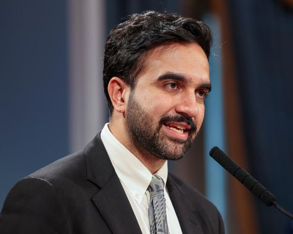
Catherine Chu, a study co-author and vice president of neurology at Kennedy Krieger and director of child neurology and pediatric epilepsy at Johns Hopkins Children's Center, framed the clinical significance directly. "We haven't understood why patients with epilepsy have problems with memory," she said, adding that the work helps close that gap.
That offers a possible explanation for something clinicians have long observed, which is that many people with epilepsy report memory difficulty despite intact daytime cognitive testing. It suggests a route toward detecting and monitoring cognitive effects of epilepsy, and eventually toward evaluating whether reducing overnight spike burden protects memory. That last step has not been tested.
What 19 Epilepsy Patients Can and Cannot Tell Us
Generalization is the central limitation and deserves stating plainly rather than in a closing line.
These were patients with drug-resistant epilepsy, a condition that changes brain networks. Many were on antiseizure medications, several of which affect sleep architecture. Some had structural abnormalities in the very regions being recorded. Electrode placement followed clinical need, so coverage differed between patients.
Whether the same hierarchy operates identically in people without epilepsy is a reasonable inference but not a demonstrated fact. Nearly all direct human evidence about deep brain activity during sleep comes from this population, which is a known constraint across the field rather than a flaw in this study.
The memory tested was motor learning, a specific type involving a practiced sequence of movements. Whether the same cascade supports factual or autobiographical memory was not examined here and should not be assumed.
For readers, nothing changes about sleep advice. The finding supports what is already recommended: adequate, consolidated non-REM sleep matters for retaining what you learned that day, and fragmented sleep interferes with it. That was known from behavioral studies. This adds the mechanism.
Anyone with epilepsy noticing memory problems should raise it with their neurologist rather than attributing it to medication or age. It is a recognized issue, and overnight EEG findings may be relevant to the discussion. Anyone with unexplained memory decline should seek evaluation, since causes including sleep apnea, medication effects, thyroid disease, and depression are treatable. This article is general information and is not medical advice.
Frequently Asked Questions
What did the study find? Slow oscillations in the orbitofrontal cortex organize spindle and ripple activity across the thalamus and hippocampus during sleep, and stronger coupling predicted better overnight memory.
Why is it the first of its kind? It is the first human study to directly link interactions among those three regions to memory consolidation, using simultaneous intracranial recordings.
Who were the participants? Nineteen patients with drug-resistant epilepsy, covering 26 sampled hemispheres, who had electrodes implanted for clinical seizure localization.
Why only epilepsy patients? Electrodes inside the brain cannot ethically be placed in healthy volunteers. This is the only routine circumstance in which such recordings exist.
Do the findings apply to everyone? Unclear. Drug-resistant epilepsy alters brain networks and medications affect sleep, so generalization is an inference rather than a demonstrated fact.
What did it show about epilepsy? Spikes coupled to sleep oscillations, especially slow oscillations, predicted worse overnight memory performance.
Does this change sleep advice? No. It supports existing guidance that consolidated non-REM sleep matters for memory, and explains part of why.