Professor Nicolas Smith, Kings College London and University of Oxford
This virtual model shows the blood flow through vessels serving the heart. During the heartbeat, different amounts of pressure are put on the different blood vessels that feed the heart – shown here in different colours. Photograph: British Heart Foundation
Dr Vanessa Ferreira, Dr Stefan Piechnik, Dr Theodoros Karamitsos and Professor Stefan Neubauer, University of Oxford
This collage of magnetic resonance imaging (MRI) scans of the heart is inspired by a new imaging technique called T1-mapping. T1-mapping uses colour to give more information about heart disease than standard black and white MRI scans. Photograph: British Heart Foundation
Professor David Greaves (University of Oxford) and Professor Ed Fisher (NYU & Eastman Visiting Professor in Oxford)
This image is a cross-section of a "fatty plaque" from a mouse artery. Fatty plaques are a mixture of "bad" LDL-cholesterol, immune cells and other material, which can build up in arteries and eventually rupture, releasing a blood clot which can cause a heart attack or stroke. This image was created using a technique called immunoflourescence microscopy. Photograph: British Heart Foundation
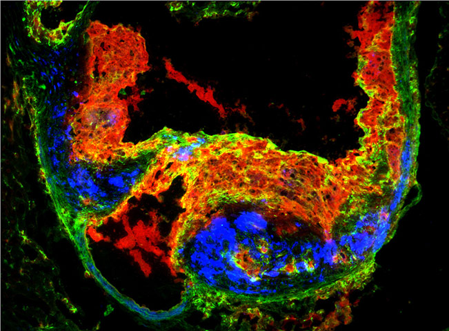
Dr Evie Maifoshie, Imperial College London
Human embryonic stem cells have the ability to turn into any cell type in the human body – including heart muscle cells. The British Heart Foundation's Mending Broken Hearts campaign, hopes to harness the potential of stem cells to grow and repair muscle which is damaged after a heart attack. Photograph: British Heart Foundation
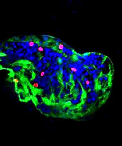
Dr Gianfranco Matrone, University of Edinburgh (BHF Centre of Research Excellence)
This image is a close-up of the heart of a zebrafish, and shows heart muscle cells in the process of dividing. Understanding the mechanisms which control the growth of heart cells in early life can give clues about how to make damaged adult hearts beat strongly again – the ultimate aim of the British Heart Foundation's Mending Broken Hearts campaign Photograph: British Heart Foundation
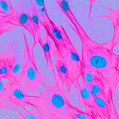
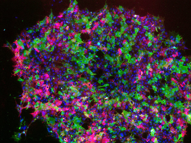
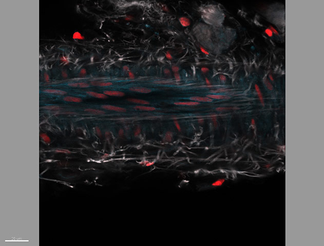
Dr Patrizia Camelliti, Imperial College London
This image shows human heart cells growing on a bioengineered "scaffold". Cells have been stained with fluorescent molecules to identify the nuclei in blue, and the cell body, in pink. The research behind this image involves working out the roles that different cells play in heart structure and function, and particularly the relationship between cells and their surrounding environment. Photograph: British Heart Foundation
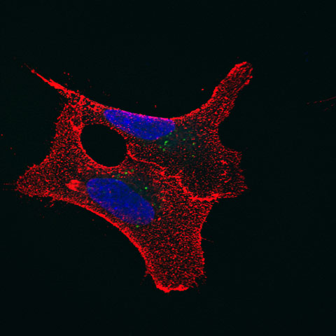
Dr Renata Gomes, Ana Lima, and Dr Ricardo Neves, University of Oxford
This image shows two blood cells which have been turned back into a stem cell-like state. The green dots are tiny nanoparticles which these researchers are using to turn these cells into heart cells and monitor them afterwards. Photograph: British Heart Foundation
Dr Richard Starke and Dr Anna Randi, Imperial College London
The inside of blood vessels are coated with a single layer of cells called endothelial cells. This image shows an endothelial cell stained to show its nucleus (purple) and the fibres that maintain its shape and allow it to move (green). A cell surface receptor, stained in red, allows the cell to bind to the vessel wall, where it can carry out its vital job of keeping blood vessels healthy. Photograph: British Heart Foundation
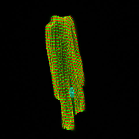
Dr Thomas Cahill, University of Oxford
This picture shows a heart muscle cell from a mouse, taken using a powerful ‘confocal’ microscope. The red and green bands show two different proteins, alpha-actinin (red) and titin (green). Alpha-actinin and titin are two of the specialised proteins in every heart muscle cell which bind and pull together to cause the cell to contract. A ripple of contraction spreads throughout the heart to cause a heartbeat. Genetic mutations in the genes which code for proteins like alpha-actinin and titin can cause inherited heart disease. Photograph: British Heart Foundation

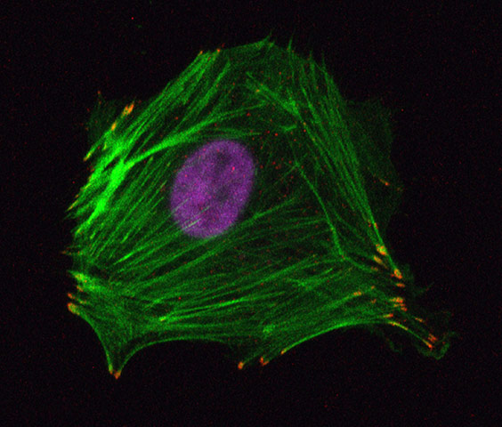
Dr Elisabeth Ehler, Kings College London
Beating heart cells – called cardiomyocytes – sit within a "scaffold" that keeps the heart in shape. Problems with this scaffold are a hallmark of some types of heart disease. This image shows a green cardiomyocyte in a Petri dish. It appears to be making contact with another type of cell, called a fibroblast, shown in red. Fibroblasts help produce the scaffold that holds the heart in shape. Understanding how these different types of cell interact will aid our understanding of how heart disease develops. Photograph: British Heart Foundation
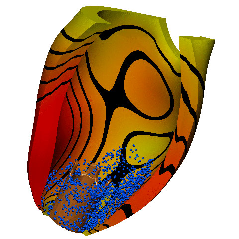
Matthew McCormick, David Nordsletten, and Professor Nicolas Smith, Kings College London
This is a computerised image of the heart of a human patient with a left ventricular assist device (LVAD) implanted into their heart to help it beat properly. The device is in the lower part of the image. This image was produced to work out how well the implanted device was working. The contours show areas of high and low pressure on the walls of the heart. Photograph: British Heart Foundation
Dr Timea Belezai, University of Oxford
This image shows a piece of leftover human coronary artery from a patient who had undergone coronary bypass surgery. This research team examined potassium channels in the vessel wall, which play a vital role in blood flow regulation. Problems with these channels can prevent arteries contracting and expanding correctly, contributing to coronary heart disease. The blue stains show calcium-activated potassium channels. The image is an extreme close-up made using a powerful confocal microscope – the red marks are the nuclei of individual cells. Photograph: British Heart Foundation