Since the past two months, students of DM Cardiology at SCTIMST, have been engaged in an exciting project, one that has given them better insight into the intricacies of the heart and the structures around it and one that promises to make them better doctors.
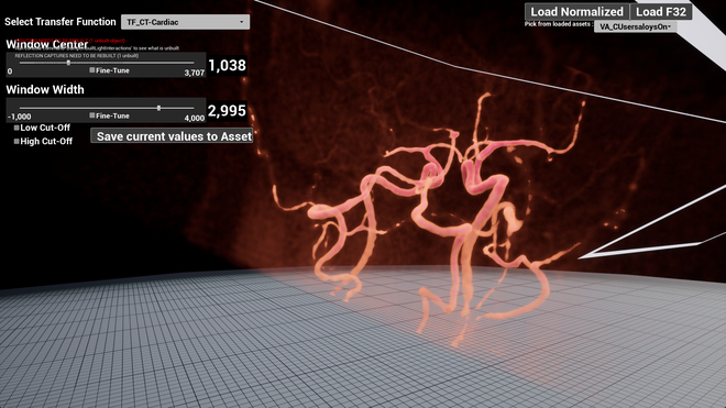
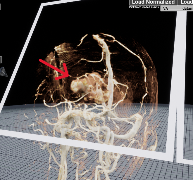
Utilising radiological images of the heart (CT and MRI scans) and using a new software as an interface, the residents have been creating a repository of 3D graphical images of the heart as is seen in various complex congenital heart diseases (CHD). They have been uploading data and case studies alongside, so that the complex anatomy of CHDs is unravelled and made simple enough for a layman to understand the difference between a normal heart and an anomalous one.
This has been made possible through a novel stereoscopic visualisation tool, one that was initially envisaged for teaching the intricacies of neuroanatomy to MBBS students .
The new tool has been developed through a collaboration between the Department of Radiology SCTIMST; the Department of Anatomy, Govt Medical College, Thiruvananthapuram and the International Institute of Information Technology (IIIT), Hyderabad
The new visualisation method utilises 3D graphic models created from MRI images of volunteers and using stereoscopic projection (the technology used in 3D movie theatres), these three dimensional figures can be projected onto a big screen for classroom teaching or a larger audience.
“Medical nomenclature and spatial orientation of the structures in the heart, particularly in CHD, is very complex. But the 3D visualisation makes the complex anatomy of a congenital heart anomaly so much easier for anyone to understand. The biggest advantage is that these images provide a more cohesive and clear picture to the cardiologist, cardiac anaesthetist, the cardiac surgeon and a cardiac radiologist who work together, and surgical planning is easier when everyone in the team speaks the same language,” Dinesh Raja P, DM Cardiology resident, SCTIMST, said.
Normal CT images are interpreted by a radiologist based on his skill and experience. But the 3D images give the surgeon or interventional radiologist a clearer idea of the different approaches they can adopt and the advantage of one over the other. The 3D images and the fact that it can be rotated or twisted and observed from various angles help doctors understand the depth of tumours or pick out something that may have been overlooked when looking through the radiological scans.
The stereoscopic visualisation of 3D images had been evolved initially to teach anatomy to MBBS students, particularly neuroanatomy, which is hard to teach and tough for students to comprehend. The idea of developing it further for use in clinical practice (surgical planning), came later
The software, which is being used as an interface to convert the DICOM images (DICOM is the international standard in radiology to transmit, store, retrieve, process and display medical imaging information) to the 3D format, was developed under the guidance of C. Kesavadas, Professor and Head, Imaging Sciences and Intervention Radiology Department, SCTIMST, by a team of students from Government Engineering College, Barton Hill (GECBH), led by Aloysious Benoy, as part of their final year project.
A tripartite MoU has also been signed between SCTIMST, GECBH and M/s Embedite Pvt. Ltd for further development of the software.
“A chunk of the anatomy lessons that are part of the MBBS curriculum has already been converted to the 3D format. But we need a wider application of the technology and more resident doctors to use this so that a repository of images from various medical specialities can be created, Dr. Kesavadas said. He added that SCTIMST & GECBH have submitted a joint patent application (No: 202341055260) for this system.
The software is affordable, platform-independent and customisable. It can seamlessly operate on any machine and can be tailored to the specific requirement of users. All it requires is a pair of overhead projectors, a computer, a screen for projecting 3D images and 3D glasses to view the images.
Anyone in the related field can easily learn the shortcut keys used in the software to rotate the image and traverse through the plane and axis.