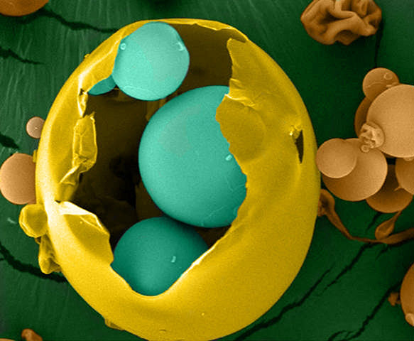
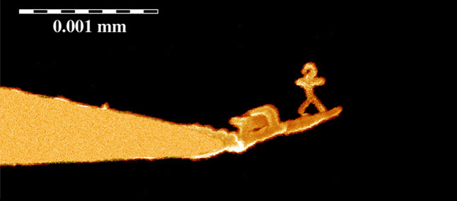
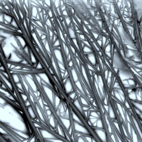
Spray drying is a commonly used technique in the pharmaceutical industry to control the particle size and distribution of powders. It typically results in shrunken and partly collapsed particles. This is due to the initial formation of a dry shell around a sphere of wet material. As the residual moisture diffuses and evaporates, the shell collapses. This image shows a small molecule compound that was spray dried to enhance its solubilityPhotograph: Shruti Gour/Drexel UniversityThis nanoman demonstrates the precision of a scanning tunnelling microscope (STM). It was created by focused electron beam deposition on the tip of a STM with a precision of 10nmPhotograph: Richard Palmer/Nanoscale Physics Research Laboratory/University of BirminghamBy controlling the assembling of peptide fibres – the building blocks of proteins – scientists have found ways to mimic the matrix material that provides structural support to cells in the body to aid growth. Such research could lead to advances in regenerative medicine. In this image, the peptides form into a highly branched structure by random assemblyPhotograph: Santanu Ray/National Physical Laboratory
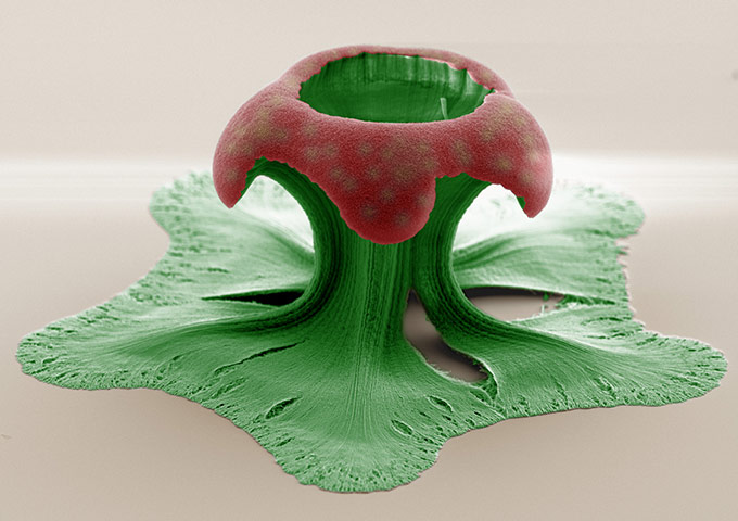
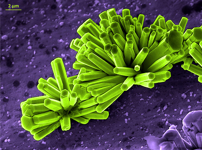
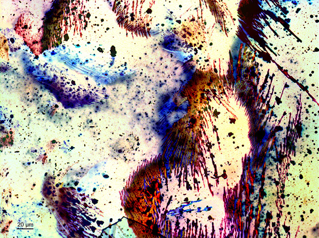
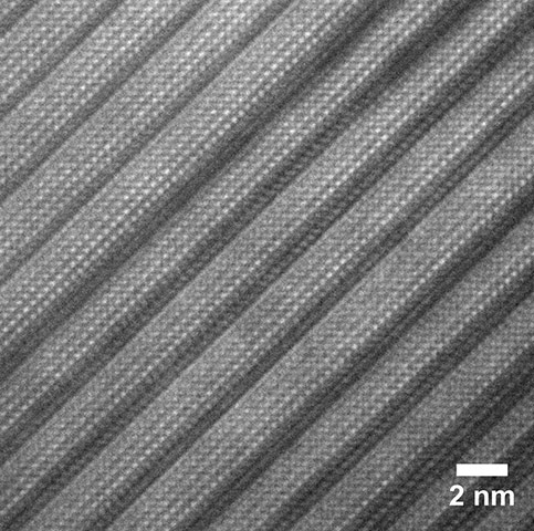
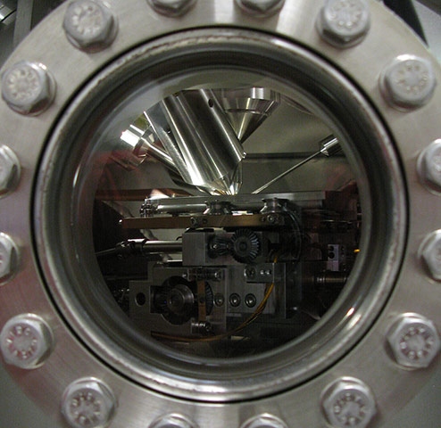
This is a colour-enhanced image of a collapsed carbon nanotube (CNT) pillar. After the pillar was formed, it was subjected to oxygen plasma treatment (to functionalise each CNT with oxygenated groups and to etch the outer portion of the pillar) and capillography process (to collapse the CNT pillars inward, increasing their packing density). This combination is necessary when the CNT pillars are used as scaffolds for microneedles. These microneedles could be used in a rapid self-administered and painless drug delivery system, replacing hypodermic needlesPhotograph: Adrianus Aria/Caltech/Materials Research Society//Science as Art CompetitionThese are one-dimensional zinc oxide (ZnO) nanorods; the high surface-to-volume ratio gives them enhanced optical and electrical properties. They are used in a range of applications in photonics and sensing. The image was captured using the Zeiss Ultra Scanning Electron Microscope in Crann (the Centre for Research on Adaptive Nanostructures and Nanodevices) at Trinity College, DublinPhotograph: Niall McEvoy/Crann/Trinity College DublinThese are magnesium sulphate crystals, known as 'epsom salts', viewed under a polarised light microscope. The striking colours are a result of interference of light travelling through the crystals. These salts are used in a variety of nano-sensors for the detection of biological molecules; they are also used to treat eclampsia in pregnant women and as an antiarrhythmic agent in cardiac arrestPhotograph: Dr Rohit Mishra/Crann/Trinity College DublinThis is a high resolution image of a material that forms crystalline grains when made by mixing chemicals in a beaker, spreading the resulting material on to a substrate and then heating the substrate to complete the reaction. The grains consist of a series of layers separated by regions where the structure of the material changes abruptly. In this image, the layers are the lighter, highly ordered regions separated by darker lines. This material is known as an Aurivillius phase material after the scientist who first observed this strange structural behaviour. At room temperature, it is simultaneously ferroelectric and ferromagnetic – unlike other examples that have to be artificially cooled to display such properties. These materials could be used in four-state memory devices based on combinations of electrical polarisation and magnetic alignmentPhotograph: Martyn Pemble/Tyndall National InstituteInside a secondary ion mass spectrometer, the surface of a material is being bombarded with ions, causing collisions that ultimately lead to the ejection of charged particles. These particles provide information on the composition of the upper few nanometres of the surface and can be mapped with sub-micron resolution. Secondary ion mass spectrometry (SIMS) is often used for the analysis of complex molecules and organics, such as biomaterials and drug delivery systemsPhotograph: Claire Hurley/Sheffield Surface Analysis CentreThis image shows keratinocytes (skin cells) migrating towards each other across a scratch. As the leading edges (lamelipodia) meet, the cells reform a cohesive sheet. Skin cells are programmed to migrate into empty space and create new epithelium when their neighbours are lost – for example, when scratched. By understanding the basic mechanics of skin regeneration, we can make advances in regenerative medicinePhotograph: Anthony Bullock/Kroto Research InstituteThese nano-mushrooms are made of nanocrystalline platinum. The stalks are only 120nm in width. These structures reveal the effect of sample size reduction on mechanical strength and fracture at the nanoscale. For mechanical testing, the mushroom head is pulled using diamond grips. Sometimes they are separated from the substrate and stick to each other in different waysPhotograph: Wendy Gu/Caltech/Art of Science
Sign up to read this article
Read news from 100’s of titles, curated specifically for you.