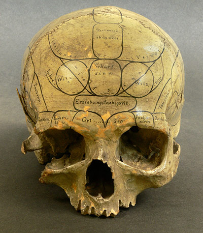
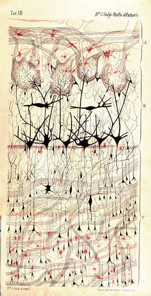
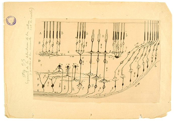
A human skull inscribed by a nineteenth-century practitioner of phrenology. According to this now discredited theory, bumps on the skull betray the volume of the brain areas beneath each one, and thus can be employed to divine a subject’s cognitive or moral strengths and weaknesses Photograph: Eszter Blahak/Semmelweis MuseumDrawing of a dog’s olfactory bulb by Italian physician and scientist Camillo Golgi. The features that appear here were revealed by a revolutionary method for staining nervous tissue that bears his namePhotograph: Camillo Golgi (1875)/Courtesy of Dr. Paolo Mazzarello, University of PaviaDrawing of the neuronal circuit found in the retina by Spanish scientist Santiago Ramón y Cajal in 1901. By applying Camillo Golgi’s tissue-staining method with patience and virtuosity, he laid the foundations for the modern field of neuroscience Photograph: Santiago Ramón y Cajal (1901)/Courtesy of Dr. Juan A. de Carlos. Cajal Legacy, Instituto Cajal (CSIC)
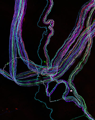
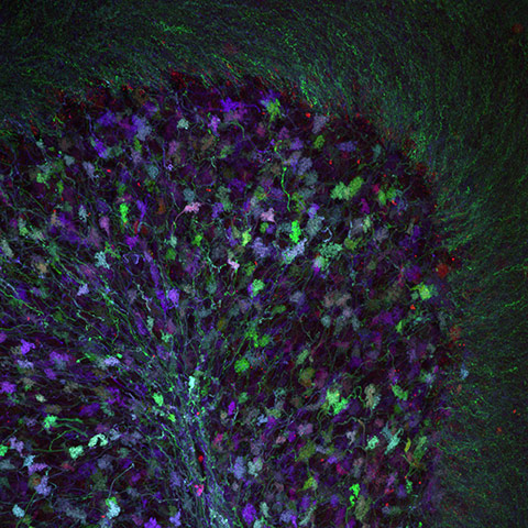
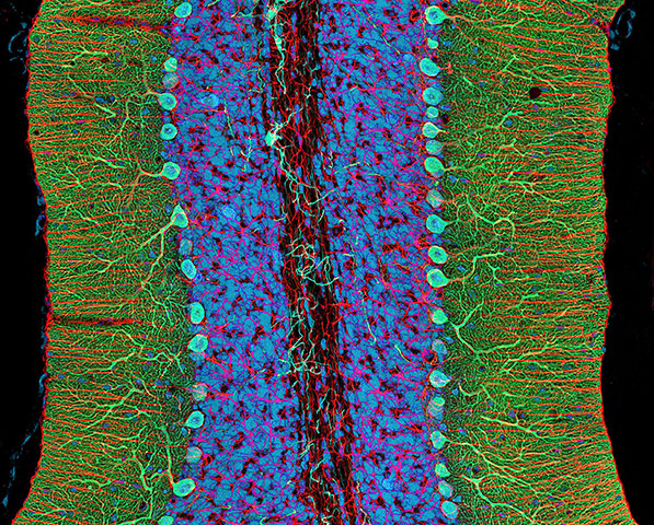
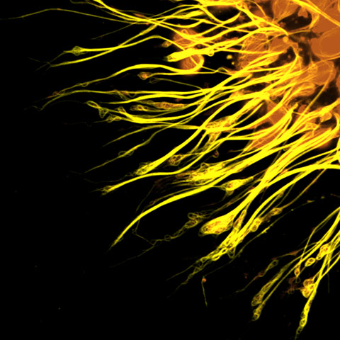
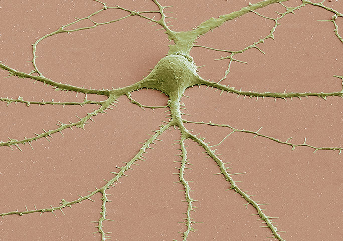
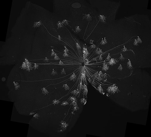
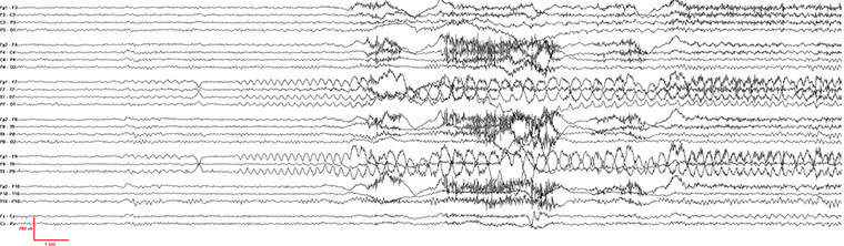
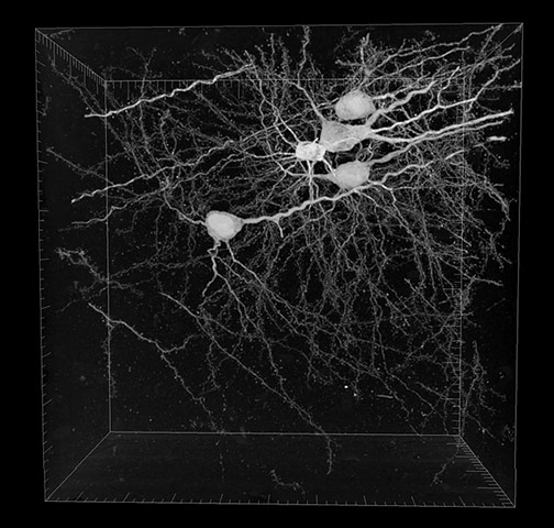
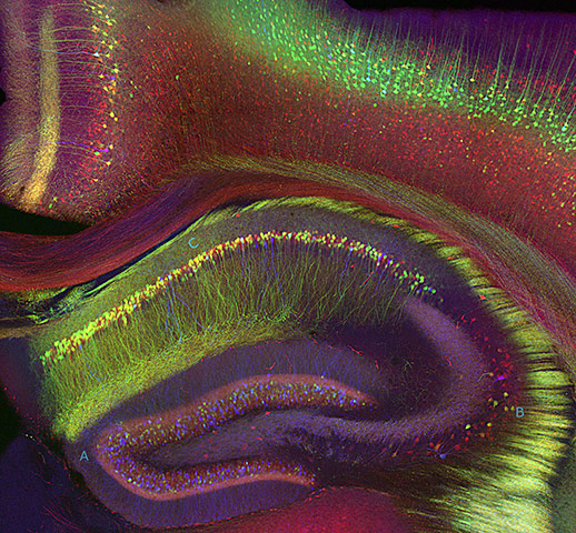
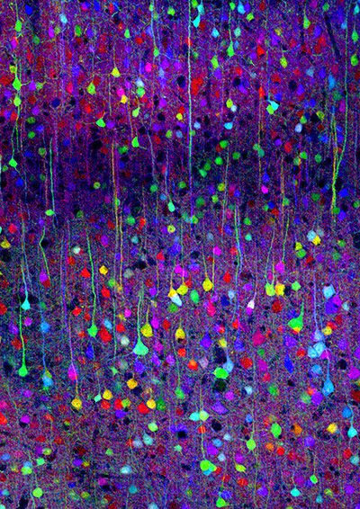
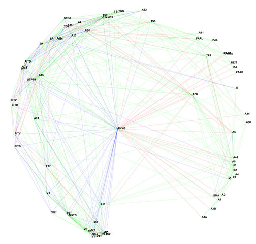
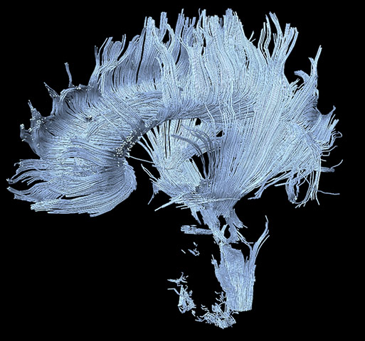
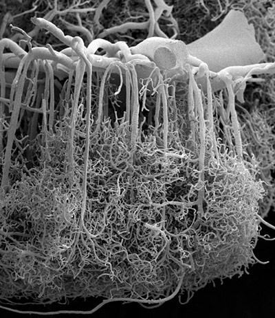
Image taken from a transgenic “Brainbow” mouse that enables neuroscientists to distinguish between neighbouring, densely packed neurons by illuminating them in different colours. This photomicrograph reveals the disposition of axons (the long portion of a neuron that generally conducts impulses away from the body of the cell) that regulate the contraction of certain musclesPhotograph: Ryan Draft, Jeff Lichtman and Joshua Sanes (2007)This photomicrograph shows presynaptic terminals in the cerebellum, which are called rosettes because of their flower-like appearancePhotograph: Tamily Weissman, Jeff Lichtman and Joshua Sanes (2007)Photomicrograph of different components of the rat cerebellum, including Purkinje neurons in green, glia (non-neuronal cells) in red, and cell nuclei in blue Photograph: Thomas Deerinck and Mark Ellisman (2004)Photomicrograph of the molecular scaffolding of axonsPhotograph: Michael Hendricks and Suresh Jesuthasan (2008)Photomicrograph of a neuron’s cell body and its dendrites radiating out of it, obtained with a scanning electron microscopePhotograph: Thomas Deerinck and Mark Ellisman (2009)A subset of neurons found in the mouse’s retina fluorescently labelled using a genetically-encoded protein. These neurons report only the motion of objects travelling in an upward direction, a feature that is predicted by the anatomy of their dendritesPhotograph: In-Jung Kim and Joshua R. Sanes (2008)Electroencephalogram of a human patient undergoing a seizure. Each horizontal line represents electrical activity recorded at a different place on the scalp. EEG recordings such as this one enable clinicians to locate the source of seizures, and guide subsequent treatmentPhotograph: Edgar Toro (2009)Five neurons automatically reconstructed by a computer from a dataset of 800 sixty-nanometer-thick consecutive sections through a small sample of nervous tissuePhotograph: Thomas Deerinck and Mark Ellisman (2009)Photomicrograph of a mouse hippocampus, an area of the brain critical for learning and memoryPhotograph: Tamily Weissman, Jeff Lichtman and Joshua Sanes (2005)Image taken from a transgenic “Brainbow” mouse that enables neuroscientists to distinguish between neighbouring, densely packed neurons by illuminating them in different colours. This photomicrograph shows a few of the many neurons that are found in the neocortexPhotograph: Tamily Weissman, Jeff Lichtman, and Joshua Sanes (2007)A summary of the known connections between the amygdala, a structure deep inside the brain that processes emotions, and the different areas of the cerebral cortex—many of which are thought to be involved in what are thought of as ‘rational’ features of the mind. The multitude of connections between these areas argues for a view in which thought and emotion are less dissociable from each other than is commonly believedPhotograph: Malcolm Young, Jack Scannell, Gully Burns and Colin Blakemore (1994)A diffusion MRI image of a patient who has suffered a stroke in the thalamus. This has resulted in major disruptions to certain axon tracts, some of which are visible at the bottom of the figurePhotograph: A Henning U. Voss and Nicholas D. Schiff (2008)Photomicrograph of the microscopic blood vessels that carry nutrients to neurons in the brain, obtained with a scanning electron microscope. This sample, from human cerebral cortex, shows a large blood vessel at the surface of the brain (top), which sends down thin, densely branched capillaries to deliver blood throughout the entire cortex Photograph: Alfonso Rodríguez-Baeza and Marisa Ortega-Sánchez (2009)
Sign up to read this article
Read news from 100’s of titles, curated specifically for you.