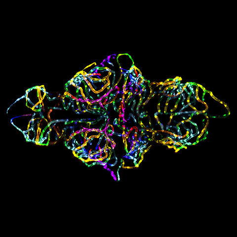
St. Jude Children's Research Hospital, Memphis, Tennessee
The blood-brain barrier in a live zebrafish embryo Photograph: Dr. Jennifer L. Peters and Dr. Michael R. Taylor
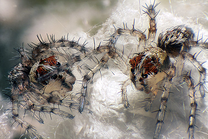
South Beloit, Illinois
Live newborn lynx spiderlings Photograph: Walter Piorkowski
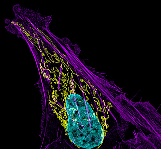
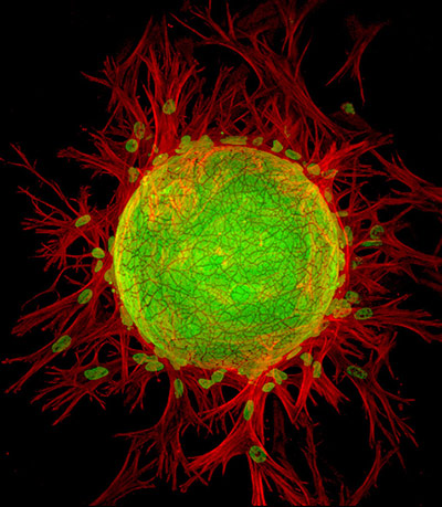
National Institutes of Health, Bethesda, Maryland
Human bone cancer showing actin filaments (purple), mitochondria (yellow), and DNA (blue) Photograph: Dr. Dylan Burnette
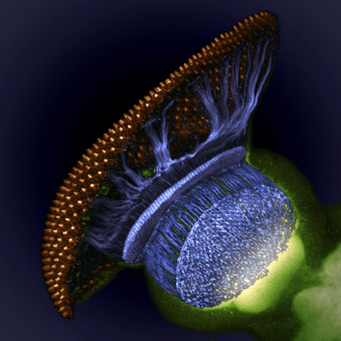
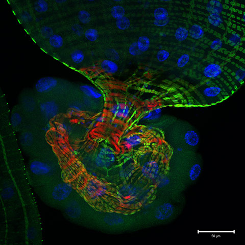
Howard Hughes Medical Institute, Ashburn, Virginia
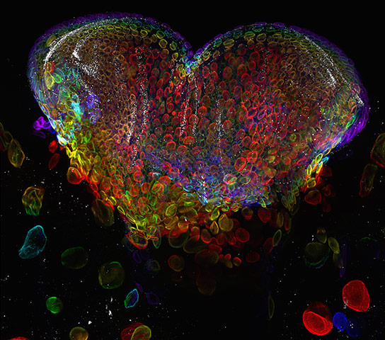
Drosophila melanogaster visual system halfway through pupal development, showing retina (gold), photoreceptor axons (blue), and brain (green) Photograph: Dr. W. Ryan Williamson
University of Valencia, Spain
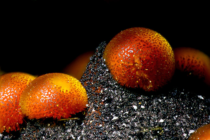
Cacoxenite (mineral) from La Paloma Mine, Spain Photograph: Honorio Cócera-La Parra
Suwalki, Poland
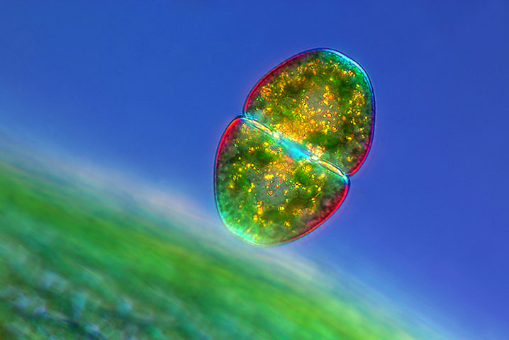
Cosmarium sp. (desmid) near a Sphagnum sp. leaf Photograph: Marek Mis
HSC Core Research Facilities - Cell Imaging Lab, University of Utah
Eye organ of a Drosophila melanogaster (fruit fly) third-instar larvae Photograph: Dr. Michael John Bridge
Düsseldorf, Germany
Pleurobrachia sp. (sea gooseberry) larva Photograph: Gerd A. Guenther
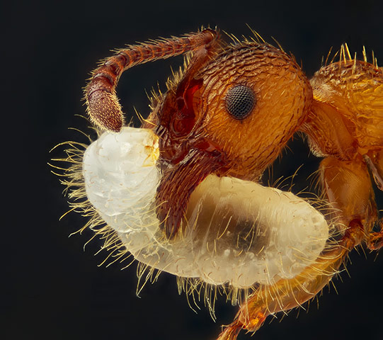
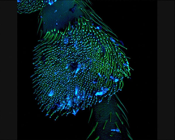
Asker, Norway
Myrmica sp. (ant) carrying its larva Photograph: Geir Drange
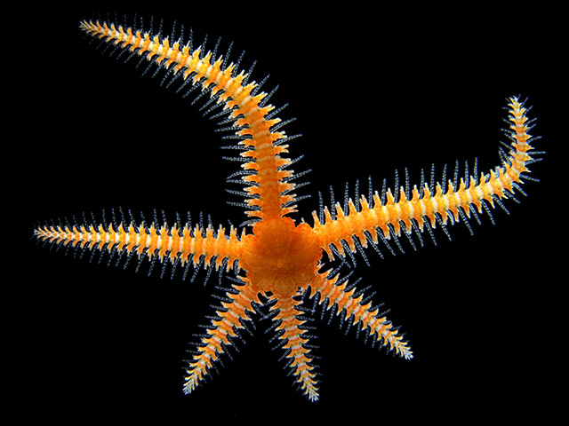
University of São Paulo, Brazil
Brittle star Photograph: Dr. Alvaro Migotto
École Polytechnique Fédérale de Lausanne, Switzerland
3D lymphangiogenesis assay. Cells sprout from dextran beads embedded in fibrin gel Photograph: Esra Guc
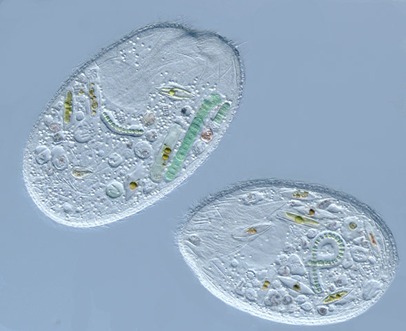
Department of Biological Sciences, George Washington University, Washington
Sonderia sp. (a ciliate that preys upon various algae, diatoms, and cyanobacteria) Photograph: Dr. Diana Lipscomb

University of Puerto Rico
Pistil of Adenium obesum Photograph: José R. Almodóvar Rivera
Department of Life Sciences and Systems, University of Turin, Italy
Section of a Coccinella (ladybug) leg Photograph: Andrea Genre
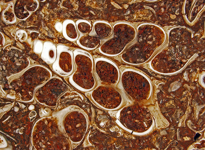
University Relations & Communications/Geology University of Wisconsin - Stevens Point, Wisconsin
Fossilized Turitella agate containing Elimia tenera (freshwater snails) and ostracods (seed shrimp) Photograph: Douglas Moore
Whitehead Institute for Biomedical Research Cambridge, Massachusetts
Single optical section through the tip of the gut of a Drosophila melanogaster larva expressing a reporter for Notch signaling pathway activity (green), and stained with cytoskeletal (red) and nuclear (blue) markers Photograph: Jessica Von Stetina
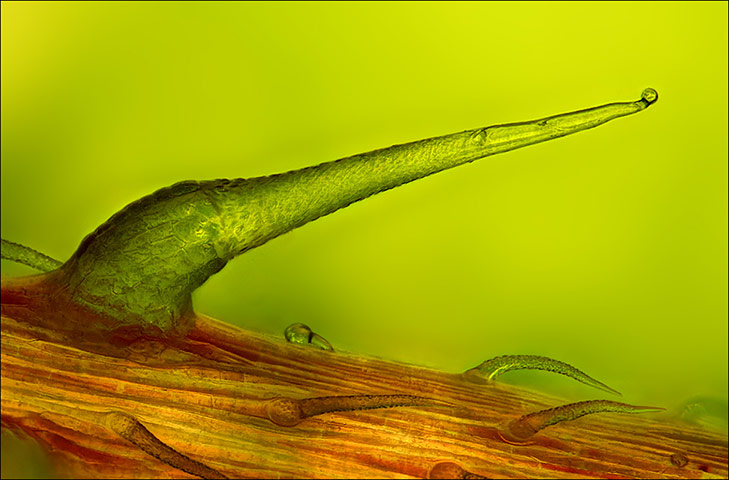
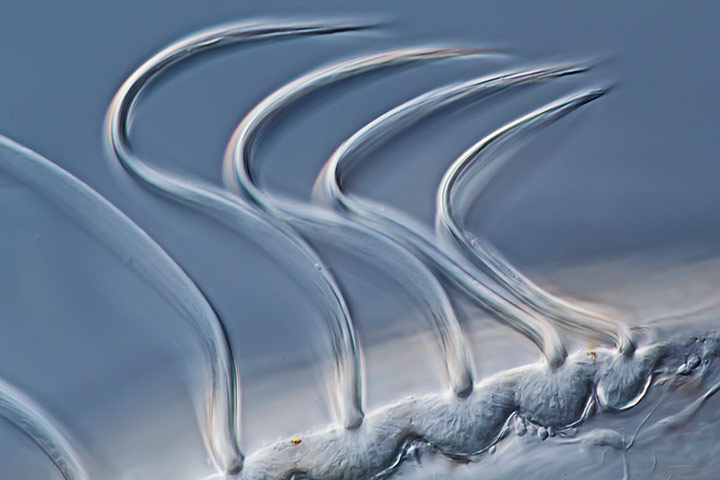
Stinging nettle trichome on leaf vein Photograph: Charles Krebs
Feltwell, UK
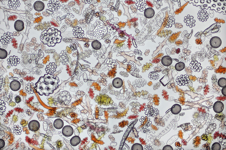
Coral sand
Photograph: Dr. David Maitland
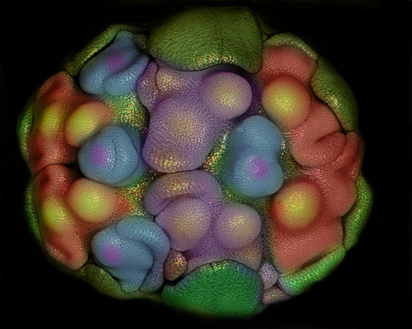
Department of Plant Biology, Faculty of Natural Sciences , University of Tabriz, Iran
Floral primordia of Allium sativum (garlic) Photograph: Dr. Somayeh Naghiloo

Department of Physiology, Development and Neuroscience, Cambridge University, UK
Embryos of the species Molossus rufus (black mastiff bat) Photograph: Dorit Hockman