Heart stoppers: winning images from British Heart Foundation competition – in pictures
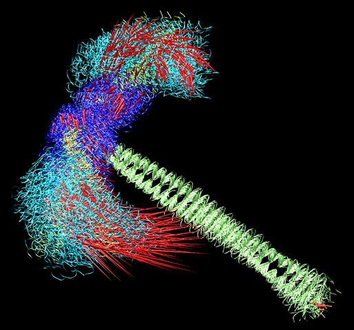
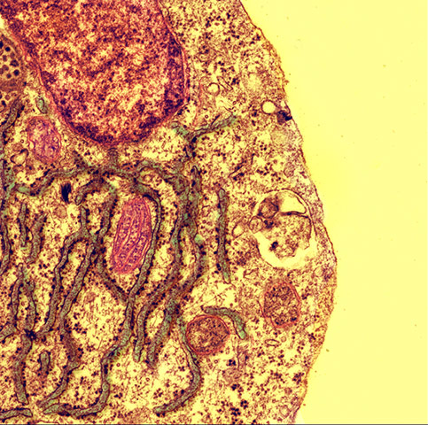
Shortlisted: Dragonfly by Arianna Fornili and Franca Fraternali at King’s College London. This image shows the simulated motion of a two-headed fragment of myosin, a molecule vital for contraction found in heart muscle cells. Different time frames of this dragonfly-shaped molecule are shown here simultaneously, with red spikes indicating the direction of motionPhotograph: Franca Fraternali/King’s College London/The British Heart FoundationShortlisted: The Forgotten Majority by Katja Gehmlich and Sina Lenski, University of Oxford. The heart is not just made up of beating cells. In fact the majority of cells are 'cardiac fibroblasts'. The internal scaffolding of fibroblasts is stained red in the picturePhotograph: Katja Gehmlich and Sina Lenski/University of Oxford/The British Heart FoundationShortlisted: Rough Road to Regeneration by Andrea Caporali, University of Bristol. This scanning-electron microscope image shows the stress response of a blood vessel cell in a condition such as diabetes. Understanding the stresses that diabetes places on the cardiovascular system may help researchers treat some of its complicationsPhotograph: Andrea Caporali/University of Bristol/The British Heart Foundation
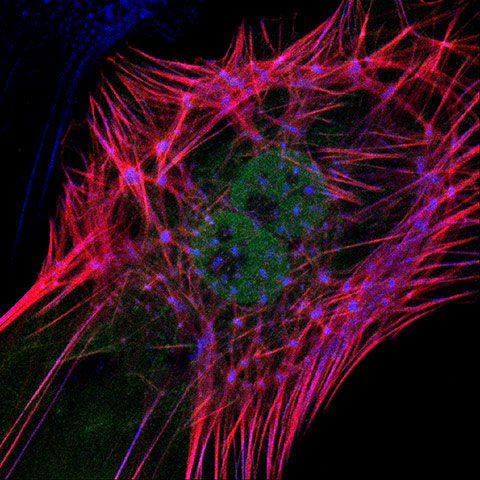
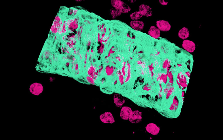
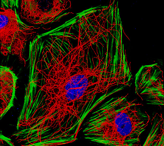
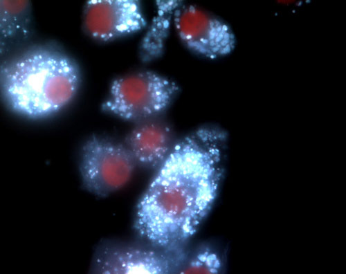
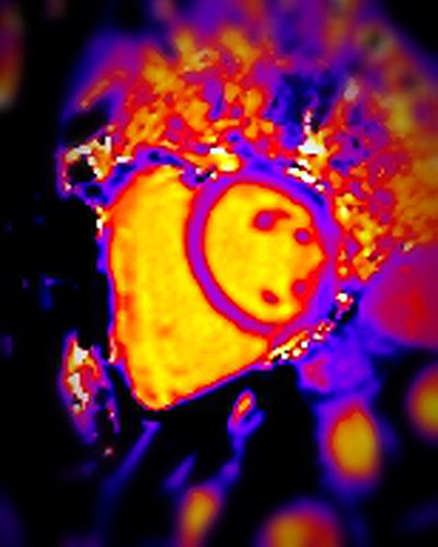
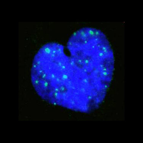
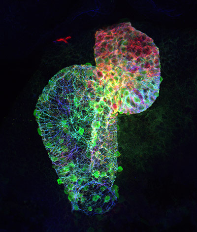
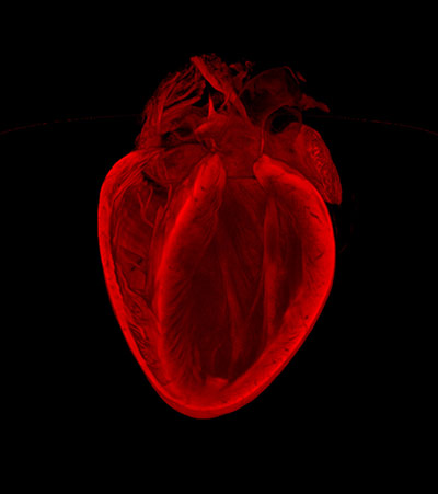
Shortlisted: At the Heart of It by Emma Kay at Queen Mary, University of London. Receptors called 'Toll-like' on white blood cells sense invading microbes and kickstart the immune response. This image shows white blood cells with Toll-like receptors (pink) migrating from a small vein (turquoise) in response to the presence of bacteriaPhotograph: Emma Kay/Queen Mary/University of London/The British Heart FoundationShortlisted: Spaghetti Junction by Graeme Birdsey and Dr Anna Randi at Imperial College London. This microscope image of cells lining blood vessels reveals the proteins that support and shape them: elongated actin stress fibres (green) and the tangled web of microtubules (red)Photograph: Graeme Birdsey and Dr Anna Randi/Imperial College London/The British Heart FoundationHighly commended: Killer Cholesterol by Dr Yichuan Wen and Dr David Leake, University of Reading. Immune cells called 'foam cells' are found in the arteries of people with atherosclerosis. The white specks are cholesterol, which can cause changes that lead to heart attacks and strokesPhotograph: Dr Yichuan Wen and Dr David Leake/University of Reading/The British Heart FoundationHighly commended: What Sets your Heart on Fire? by Dr William Moody, University of Birmingham. This is an MRI scan of the heart of a healthy kidney donor. MRI is sensitive enough to detect early, treatable scar formation in the heart that may occur after donating a kidneyPhotograph: Dr William Moody/University of Birmingham/The British Heart FoundationHighly commended: At the Heart of a Cell by Dr Andrew Cobb, King’s College London. A heart-shaped nucleus in a cell from vascular smooth muscle, which helps give blood vessels their shape. The green specks show regions of DNA damage, which could explain the unusual shape of the nucleusPhotograph: Dr Andrew Cobb/King’s College London/The British Heart FoundationMending Broken Hearts winner: Caught in the Net by Dr Jana Koth, University of Oxford. This is the developing heart of a two-day old zebrafish embryo. The green cells are heart muscle cells, and the red and blue show components that make up the muscle. The early heart tube has started to loop. It consists of two sections: the large, thin atrium (where blood flows in) and the smaller, thicker ventricle (where blood leaves flows out)Photograph: Dr Jana Koth/University of Oxford/The British Heart FoundationImage of the Year: The Broken Heart by Gillian Gray, Megan Swim and Harris Morrison, University of Edinburgh. This image reveals the 3D structure of an adult mouse heart. It was created using a technique called 'optical projection tomography', which can be used to assess the extent of injury and healing after a heart attackPhotograph: Dr Gillian Gray, Megan Swim and Harris Morrison/University of Edinburgh/The British Heart Foundation
Sign up to read this article
Read news from 100’s of titles, curated specifically for you.


.jpg?w=600)