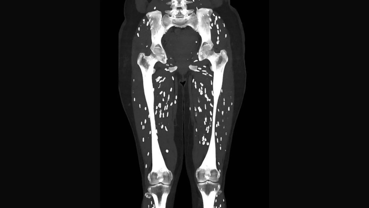
The CT scan image of a patient is going viral over the internet. In the image we see tiny spots in the legs of the individual and you would be shocked to know that these tiny spots are the tapeworm cysts.
The image has been shared by Sam Ghali on X (formerly Twitter). "Here’s one of the craziest CT scans I’ve ever seen, What’s the diagnosis?," he captioned the picture.
In subsequent posts made on X, he has explained that it is a condition called cysticercosis which is caused when one consumes larval cysts of Taenia Solium or tapeworm.
Cysticercosis is a parasitic infection caused by the larval stage of the tapeworm Taenia solium. It occurs when individuals ingest the eggs of this tapeworm, typically through contaminated food or water. Once inside the body, the eggs hatch into larvae that migrate to various tissues, forming cysts. This condition commonly affects muscles, eyes, and the brain.
In the brain, cysticercosis can lead to neurocysticercosis, a serious form of the disease that causes neurological symptoms such as seizures, headaches, and confusion. In the muscles, it might present as pain or swelling. Diagnosis often involves imaging techniques like MRI or CT scans, which can reveal the cysts.
The expert explains the lifecycle of the tapeworm larval cyst. "So humans become infected with T. Solium by ingesting cysts that can be found in undercooked pork. After several weeks (usually around 5-12) these cysts evolve within the gastrointestinal tract into mature adult tapeworms. This condition is known as Intestinal Taeniasis.
These adult tapeworms then shed eggs which are in turn excreted into human feces. It’s very important to note that it is only when these eggs are ingested via fecal-oral transmission, that one can develop the clinical syndrome known as of Cysticercosis!," he explains in the long post.
After the eggs are ingested (humans or pigs) they release larvae which penetrate the intestinal wall and invade into the bloodstream (via mesenteric venules) and from there can spread to literally anywhere in the entire body. The brain, eyes, subcutaneous tissues, and skeletal muscles are the most common destinations. The larvae lodge wherever the end up and ultimately form cysts known as cysticerci, he adds.
Is the human body capable of eliminating them?
Yes! In many cases, the immune system can partially or fully eliminate cysts over time, particularly if the infection is limited to the muscles. The cysts in these areas might be gradually destroyed and reabsorbed by the body.
"The hosts inflammatory response typically ends up killing off the cysts, which subsequently undergo calcification, giving them the classic appearance you can appreciate on this CT scan. These are commonly referred to as “rice grain calcifications”, Sam Ghali explains.
However, when cysticercosis affects the brain, known as neurocysticercosis, the situation is more complex. The immune system's response to the cysts in the brain can cause inflammation and potentially lead to neurological symptoms such as seizures or headaches. "The prognosis for cysticercosis is generally good but unfortunately some cases are fatal. It's estimated that around 50 million people worldwide are infected each year resulting in ~50,000 deaths," he adds.